Showing posts with label dissection. Show all posts

Friday, July 20, 2012

"Dissection on Display: Cadavers, Anatomists and Public Spectacle" by Christine Quigley, Book Review by Bess Lovejoy

I am very excited to read the new and wonderful looking book Dissection on Display: Cadavers, Anatomists and Public Spectacle by Christine Quigley, who many of you might best know as editor of the wide-ranging blog Quigley's Cabinet. I have not yet had a chance to read the book, but we are all in luck, as friend of Morbid Anatomy Bess Lovejoy--author of the forthcoming Rest in Pieces: The Curious Fates of Famous Corpses--has kindly offered up a very detailed and thoughful review of the book special for the Morbid Anatomy blog.

Lovejoy's review follows; you can also find out more about the book--or purchase a copy of your very own--by clicking here.
Dissection on Display: Cadavers, Anatomists and Public Spectacle
By Christine Quigley
Mcfarland, February 24, 2012

Most of us have never seen a dead body, let alone witnessed the dissection of a cadaver. But for centuries in Europe, Britain, and America, public dissections were highly social occasions. In the candlelit, damask-draped anatomy theatre of 18th century Bologna, townspeople jostled medical students and high-ranking officials during a two-week-long dissection that took place as part of the annual carnival. In 16th century Britain, hundreds crowded around to watch the dissections of executed criminals. And in early 19th century America, the most fashionable strata of society (men and women alike) attended public dissections for a chance to “see and be seen.”
In her new book, Dissection on Display: Cadavers, Anatomists and Public Spectacle, scholar, author, and blogger Christine Quigley traces the hidden history of anatomists who perform for the public. Not all of the men she profiles are known just for their dissections: some, like 17th and 18th century Europeans Frederik Ruysch and Honoré Fragonard, did the dirty work in private, then displayed the exquisitely-crafted results to the public in the form of art and illustration. Many of the names Quigley profiles will be familiar to Morbid Anatomy readers, though others -- like Thomas “Mummy” Pettigrew, the 19th century London antiquarian who unrolled mummies to entertain his guests – may be fresh discoveries.

Using a series of thematically-grouped vignettes, Quigley explores anatomists as demonstrators, educators, collectors, showmen, and more. Some of the book’s most intriguing passages deal with the lessons that public dissections were supposed to impart: not just about the workings of the body, but the workings of God, and of justice. Even more than a chance to gain medical wisdom, public dissections were often promoted as an opportunity to witness the glory of God in the functioning of a corpse’s entrails. Sometimes they were also seen as a chance to exult in the final stage of  punishment meted out to a criminal. The mutilation of the corpse was thought to deny the deceased a chance at Resurrection -- thus condemning him or her both in life and afterlife.

Quigley also touches on the racial and sexual undertones that have long troubled the study of anatomy. One of the book’s most disturbing sections profiles French naturalist and zoologist Georges Cuvier and his quest to uncover (literally) the mysteries that lay between the legs of Saartjie Bjartmaan, also known as the “Hottentot Venus.” Bjartmaan – a young Khoisan woman from South Africa -- entertained audiences in early 19th century London and Paris with the enormous size of her buttocks. Some whispered that Bjartmaan was also blessed with a similarly enormous labia minora, and like other scientists of the time, Cuvier was fascinated by such rumors. After Bjartmaan’s death, he detailed her dissection in a medical journal and preserved both her brain and genitals in greenish glass bottles outside his office. Thus the last shred of modesty that Bjartmaan had protected in life was unceremoniously stripped from her in death, in a way that calls to mind the brutally frank autopsy reports of modern dead celebrities.

Today, human dissection is usually hidden from the public. This cloaking began in the 19th century, when, as Quigley writes, “The anatomists withdrew behind the doors of educational institutions, and the townspeople were not invited to join them.” These days dissections occur exclusively in a medical or forensic context, and the only corpses we see are on television. No longer is the public treated to theatrical displays of their own inner-workings, as they were in the days when Andreas Vesalius kept Renaissance audiences glued to their seats.

But there have been exceptions. In 2002, the controversial Gunter von Hagens – he of the plastinated corpses and Body Worlds exhibits – staged a ticketed dissection of the body of a 72-year-old man in London. The event drew considerable attention, and Hagens faced the threat of arrest even while wielding the scalpel. Yet the room was packed, proving that our appetites for dissection haven’t waned. Quigley includes an excellent photograph of the event, notable not for the pale cadaver about to be sliced apart, but for the front row of the audience, their faces horrified, bemused, and fascinated in turn. One woman crosses her hands over her chest in protection, clutching her check and beginning to grimace. Next to her, an older gentleman folds his wrists behind his elbows and leans back as if to say “show me what you got.” Von Hagens himself is at the forefront of the image, clad in a black fedora -- his nod to Rembrandt’s “The Anatomy Lesson of Dr. Nicolaes Tulp,” which hangs on the wall above him.

In fact, Quigley takes pains to show us how conscious Von Hagens – the most famous modern anatomist -- is of his historical lineage. (Many of his most famous pieces, such as his flayed horse and rider, quote directly from the work of earlier anatomists such as Fragonard.) This is where the book shines: Quigley has stitched together a family tree of public anatomists who contributed to our understanding of the body, but whose work often remains hidden like the organs beneath our skin. Dissection on Display is recommended reading for anyone with a healthy sense of curiosity, morbid or otherwise, about what used to happen when we were allowed to watch.
The writer of this post, Bess Lovejoy, is a writer, editor, and researcher based in Seattle. Her book Rest in Pieces: The Curious Fates of Famous Corpses is coming out March 2013. You can find out more about her at her website besslovejoy.com." To find out more--or to purchase a copy of this book--click here.

Image: The Anatomy lesson of Dr Nicolaes Tulp, Remrandt van Rijn, 1632; found on Wikipedia. According to Quigley, the dissection was performed in Leiden’s anatomical theatre, and included an audience of townspeople that were left out of the painting. Instead, Rembrandt was paid to include surgeons who may or may not have actually been there.

Wednesday, July 18, 2012

"Dissection as Studio Practice with Real Anatomical Specimens," Class with Laura Splan, Observatory, Next Monday, July 23

I am very excited about "Dissection as Studio Practice," a class taking place next Monday, July 23, at Observatory. Taught by artist Laura Splan, the class will begin with an illustrated survey of the use of notions of "dissection" in contemporary art practice; these principles will then be applied to in-class projects which include the dissection of your very own anatomical specimens (i.e. frog, sheep brain, cow eye). This class is open to all expernience levels, and participants are invited to bring additional materials, objects and artifacts that will inspire their “dissective” inclinations.

We had a wonderful time in the last iteration of this class, as you can see from the photos above. Full description of the class follows. Class size is limited; if interested, be sure to RSVP via email to morbidanatomy[at]gmail.com. You can see more of Laura Splan's work by clicking here. Hope very much to see you there!
Lecture and Studio Art and Dissection Class with artist Laura Splan
Date: Monday, July 23
Time: 7-10 PM
Fee: $75
*** Class size is limited to 16; please RSVP to morbidanatomy[at]gmail.com

This class is part of the
Morbid Anatomy Art Academy

This class will survey the use of dissection in contemporary art practice through an illustrated lecture, specimen dissections, and studio time for individual and collaborative projects. We will examine the conceptual and cultural significance of cutting, excavating, disassembling, labeling, observing and displaying “bodies.” The lecture will present a brief history of dissection as well as work by contemporary artists exploring imagery, tropes and methods of dissection. The collaborative and individual art projects will be fun and lively hands on explorations of the meaning of dissection in a work of art. Each student will receive a complete specimen dissection kit (i.e. frog, sheep brain, cow eye) to create a self-directed dissection project with. Participants should bring additional materials, objects and artifacts that will inspire their “dissective” inclinations. Additional supplies will be provided by the instructor to stimulate your creativity. No prior art training is required. Everyone is welcome.

Laura Splan is a Brooklyn based visual artist. Her mixed media work explores historical and cultural ambivalence towards the human body. She was a Visiting Lecturer at Stanford University where she taught “Art and Biology”. She has been a Visiting Artist at the New York Academy of Sciences, California College of Art, San Francisco Art Institute, Maryland Institute College of Art, and Cal Arts. Her artwork was recently commissioned by the CDC Foundation. She curates the visual portal DomesticatedViscera.com. Images of her artwork can be found on her website: LauraSplan.com. You can find out more here. Feel free to contact Laura through her website with any questions about the class by clicking here. You can see photos from the last class by clicking here.
You can find out more here, and RSVP with an email to morbidanatomy[at]gmail.com.

Tuesday, February 21, 2012

"Surgery" Worrydoll, Mixed Media, Renée Laferriere Cinderhouse, 2011


"Surgery," Stoneware, Mixed Media, 2011. From the series Worryldoll by Renée Laferriere Cinderhouse. You can see more of her work and find out more here.

Found here.

Saturday, January 14, 2012

Philippe Verheyen Dissecting His Amputated Limb, ca. 1715


Philippe Verheyen (1648-1711) Dissecting His Amputated Limb

By an anonymous artist, ca. 1715. Postmortem painting in honor of a famous Dutch anatomist and surgeon.

Medium: Oil on Panel. Size: 16.3”X16.5”

From the collection of Pieter Deheijde.
Image and text via 100% Spleen.

Monday, November 28, 2011

"Dissection as Studio Practice" Illustrated Lecture and Studio Art Class with Artist Laura Splan at Observatory


I am super excited to be announcing the upcoming class "Dissection as Studio Practice" at Observatory on Sunday, January 8th. I met the teacher--Laura Splan--at a conference many years back now. Since then, I have been a big fan of her work, a few examples of which can be seen above, including--top to bottom-- an installation view of her current solo show Reformulations; a blood-on-watercolor composition entitled "Elaborative Encoding"; and 3 images from her "Doilies"series of 2004, a set of computer machine embroidered doilies with the design of each doily based on a different viral structure; pictured here, top to bottom: Herpes, Sars, and Influenza.

This class--open to all experience levels--will "survey the use of dissection in contemporary art practice through an illustrated lecture, discussion and collaborative art project"; it will also provide a terrific opportunity to work with an accomplished and sophisticated conceptual artist while gaining insight into process and method behind the creation of iconic and powerful works dealing with dissection and the body. I, for one, simply cannot wait!

Full description of the class follows. Class size is limited; if interested, be sure to RSVP via email to morbidanatomy[at]gmail.com. You can see more of Laura Splan's work by clicking here. Hope very much to see you there!
CLASS: Dissection as Studio Practice
Lecture and Studio Art Class with artist Laura Splan
Date: Sunday, January 8th
Time: 1-4 PM
Fee: $60
*** Class size is limited to 20; please RSVP to morbidanatomy[at]gmail.com

This class will survey the use of dissection in contemporary art practice through an illustrated lecture, discussion and collaborative art project. We will examine the conceptual and cultural significance of cutting, excavating, disassembling, labeling, observing and displaying “bodies.” The lecture will present a brief history of dissection as well as work by contemporary artists exploring imagery, tropes and methods of dissection. The collaborative project will be a fun and lively hands on exploration of the meaning of dissection in a work of art. Participants should bring an object, artifact or specimen to “dissect” for the group exercise. Additional supplies, tools and materials will be provided. No prior art training is required.

Laura Splan is a Brooklyn based visual artist. Her mixed media work explores historical and cultural ambivalence towards the human body. She was recently a Visiting Lecturer at Stanford University where she taught “Art and Biology” in the Art & Art History Department. She has been a Visiting Artist at the New York Academy of Sciences, California College of Art, San Francisco Art Institute, Maryland Institute College of Art, and Cal Arts. She curates the visual portal DomesticatedViscera.com. Images of her artwork can be found on her website: LauraSplan.com.

You can contact Laura through her website with any questions about the class by clicking here.

If you are interested in signing up for this class, please email me at morbidanatomy[at]gmail.com. To see more of Laura Splan's fantastic work, click here. This class is one of the newest installments in the series newly termed The Morbid Anatomy Artist Academy; to find out more about that--including a full class list thus far--click here.

Monday, October 3, 2011

Morbid Anatomy Presents This Week and Beyond at Observatory


Solitary vice? Sex and dissection in Georgian London
An illustrated lecture with Dr. Simon Chaplin of the Wellcome Library, formerly of the Hunterian Museum
Date: This Tuesday, October 4
Time: 8:00 PM
Admission: $5
Presented by Morbid Anatomy

In his watercolour of a 'Persevering Surgeon', the British artist Thomas Rowlandson made no bones about the darkly erotic nature of anatomical dissection. Poised over the body of a naked woman, erect knife in hand, Rowlandson's anatomist conjured images of the other solitary vice that consumed later 18th century moralists and medical men. But like Rowlandson - who combined popular satirical illustration with a more discreet trade in pornographic imagery - anatomists maintained a delicate balance between personal pursuits and public propriety. In this lavishly illustrated lecture, Simon Chaplin explores the sexual undertones of the anatomy schools of Georgian London, in which students dissected grave-robbed bodies in the back-rooms of their teachers' houses, while their masters explored new strategies for presenting their work to polite audiences through museums and lectures.

Dr Simon Chaplin is Head of the Wellcome Library in London. Before joining the Wellcome he was Director of the Hunterian Museum in London, one of the world's oldest anatomy collections. His research interests include the history of anatomy, surgery and museums, and his doctoral thesis explored the relationship between dissection and display through the work of the Hunterian Museum's founder, the surgeon John Hunter (1728-1793).

Image: Thomas Rowlandson, 'The Persevering Surgeon', late 18th century, from the collection of the Hunterian Museum, London

Born of the Floating World: A Brief Exploration of the Japanese Graphic Narrative
Illustrated talk with Japanese Scholar Dev Avidon
Date: This Thursday, October 6th
Time: 8:00 PM
Admission: $5
Presented by Morbid Anatomy and Oxberry Pegs

In 1988, the anime adaptation of Otomo Katsuhiro's perennial serialized manga Akira was released in Japan, shattering domestic attendance records for an animated film. Shortly thereafter, its distributor presented it to George Lucas and Steven Spielberg as the ideal anime for English-language adaptation. The two dismissed it offhand, deeming the very concept of a cerebral, macabre, challenging animated film as “completely unmarketable” in the United States. Animation in America was the sole purview of children, they reckoned, and throughout the 90s, those few anime that were adapted for the American market were heavily edited to remove any and all controversy or 'adult' themes, thus rendering them safe for Saturday mornings. In 2001, however, Disney took a chance, bringing a largely faithful, unedited translation of Hayao Miyazaki's Spirited Away to theaters. It was an astounding success, enjoying uniform critical acclaim and record profits for a foreign animated film, and sparking an unprecedented interest in anime and manga in America.

A decade later, with anime and manga's popularity in the United States at an all time high, Dev Avidon will present a brief history and overview of the two forms, and examine their provenance in 17th and 18th century Japanese pictorial narratives such as the Kibyoshi – arguably the first graphic novel in human history. What spawned anime and manga as media, and what defines them? How did their visual and narrative tropes and themes evolve from both a Japanese cultural tradition dating back as early as the 9th century, and a cross-pollination and interplay with Western art and animation styles during the 19th and 20th centuries? And how can we, as American viewers, reconcile our preconceptions of the 'cartoon' and 'comic book' with the realities of two artistic forms that, collectively, account for over half of all visual media produced in Japan annually?

Dev Avidon is a Brooklyn-based jazz composer/singer, sound engineer, small business owner and Japanese scholar. During his time in the Government and East Asian Languages and Civilizations departments at Harvard University he specialized in Japanese neo-nationalism and Japanese popular art, from the ukiyo-e and kibyoshi forms of the Edo period through the modern day Neopop and Superflat movements. His thesis for the Reischauer Institute of Japanese Studies at Harvard University, Allegories of the Empty Center: A study of Modern Japanese Nationalism explores how Japanese neo-nationalism as exemplified in cultural movements such as Superflat problematizes the core tenets of modern nationalist theory. He also played an instrumental role in the founding and cataloging of the anime and manga collections at the Harvard Yenching Library. A well-published author and scholar, Dev Avidon's current focus is on his upcoming sophomore album, Tears of Men, and melding the hard science of audio technology with the clean lines of Japanese aesthetic design and the artwork of master jewelers with his line of audiophile cables for Frost Audio, a boutique audio company he owns and co-founded in 2010. Visit him at www.devavidon.com or www.frost-audio.com.

Image: Hokusai's Manga

The Rest of October:

NOVEMBER:

You can find out more--and get directions to Observatory--by clicking here.

Tuesday, November 30, 2010

"The Manner of Dissecting the Pestilentiall Body," 1666


"The Manner of Dissecting the Pestilentiall Body," 1666.

Click on image to see larger, more detailed version.

Via The William Eamon blog.

Saturday, August 28, 2010

Another Take on the Anatomist and his Beautiful Cadaver, Circa 1920


"An Anatomy Lesson," ca. 1920, Vintage gelatin silver print.

Click on image to see larger, finer version.

Found on the Joseph Bellows Gallery webpage, via Bits and Bites Tumblr.

Friday, May 7, 2010

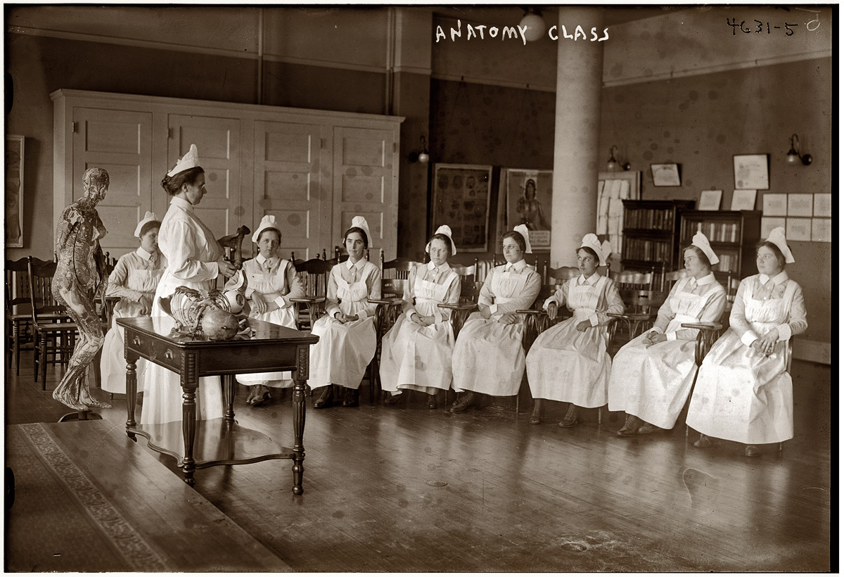
Anatomy Class featuring Auzoux Female Anatomical Model, George Grantham Bain Collection, Shorpy


Anatomy Class circa 1905, George Grantham Bain Collection, as found on Shorpy. Click on the image to see much larger, more detailed image; note especially the demure Auzoux female anatomical model to the left; you can see a color version of it here.

More on Auzoux and his work here; Via Turn of the Century blog.

Monday, April 26, 2010

"Anatomical Art: Dissection to Illustration," Exhibition Curated by Marie Dauenheimer, Arlington, Virginia


"We have the gift of language, the gift of making memories in words and pictures," Aziz says. "The bodies are gone, but by making these memories, we, to an extent, resurrect them. This is the antidote to death."--David Montgomery, "In gross anatomy, Howard U.'s Ashraf Aziz sees nothing but grace," Washington Post, Wednesday, April 21, 2010
Marie Dauenheimer, friend, friend of Morbid Anatomy, and Observatory lecturer past and future (more on that future lecture--which is entitled "Three Unique Medical Museums in Northern Italy" and will take place on Saturday May 1--soon!) has curated an exhibition of artworks inspired by the cadaver as part of a year-long collaboration between Howard's College of Medicine and the Art Institute of Washington. The Washington Post just ran a really lovely and in-depth story about the exhibition and the collaboration; it was such an interesting article that I have posted it here in its entirety:
In gross anatomy, Howard U.'s Ashraf Aziz sees nothing but grace
By David Montgomery
Washington Post Staff Writer
Wednesday, April 21, 2010

At 71, Ashraf Aziz has spent half his life cutting open cadavers and initiating medical students at Howard University into the pungent mysteries of human anatomy. He likes the heads. Extracting the muscles of chewing is one of his specialties. There is little about the body that can faze him anymore.

And yet, here in a little art gallery across the street from the Rosslyn Metro station, this most eloquent and loquacious man of science has been rendered almost speechless.

"I, I'm really a little bit -- " he stammers.

He sounds like a first-year medical student about to be overcome in the embalming room.

"I'm sorry," he says.

Tears well in his eyes.

Surrounded by red-wine-sipping artists and art students on a recent evening, the anatomist is contemplating their renderings of the human body. The nudes not only have no clothes, they also have no skin. The subjects are cadavers -- cadavers from the lab where Aziz teaches gross anatomy.

"I'm so deeply moved," he says. He never thought he would find such kindred spirits.

This exhibit, "Anatomical Art: Dissection to Illustration," is just one outcome of an unusual year-old collaboration between Howard's College of Medicine and the Art Institute of Washington. The cross-disciplinary ping-pong continues strong in both directions:

There is talk of enlisting animators from the art school to enhance lessons on locomotion at the medical school. An Art Institute sculptor presented Aziz with some fancy modeling clay, which he used to take an impression from the inside of a chimpanzee skull as part of his comparative chew-muscle research.

Meanwhile, at the art school, to such curriculum standards as "life drawing" -- portraits of live, nude models -- has been added the informal option of, well, dead drawing, in the cadaver lab. Some artists have taken to exploring -- and memorizing the Latin names of -- complex muscle groups that invisibly influence the supple motions of the living torso.

Perhaps most striking has been seeing the life and career of one of Washington's singularly passionate scientists come full circle.

The son of amateur artists in the Indian diaspora, growing up in Tanzania, Aziz, too, once wanted to become an artist. But his poet-carpenter father and his henna-painter mother feared there wasn't a secure future in art. So Aziz became a zoologist and a human anatomist, an associate professor at Howard.

The choice was apt, for the study of anatomy has embraced art -- and vice versa -- at least since Leonardo da Vinci performed dissections and drew the results. From "Gray's Anatomy," the seminal 19th-century illustrated text, to latter-day sensational exhibitions of plasticized cadavers such as "Bodies" and "Body Worlds," the dissected corpse continues to fascinate, repulse, instruct and inspire.

Aziz believes that when science and art diverge, both lose. He draws his own illustrations. To conclude his regular medical lecture on the hand and forearm, he has had traditional Indian tabla drummers demonstrate the dexterity of the muscles and tendons that his students will tweeze in the cadaver lab. In 1999 he co-wrote a 19-page academic journal article in which he cited Virginia Woolf, Gertrude Stein, James Joyce and Marshall McLuhan, along with medical authorities. The ostensible subject was to defend the relevance of the "real" cadaver in medical education despite the rise of digital "cyber cadavers" -- but really the piece was a stirring meditation on authenticity. Away from the lab he reads poetry and writes essays on popular Indian cinema.

Aziz's dream has been to see what a new generation of artists would bring to a new generation of cadavers. Even if the results were nothing revolutionary, he thought, the process itself would be illuminating for all involved: Where life has ended, insight might begin.

Anatomy for art students
As Aziz was casting his thoughts from the realm of science to art, an artist named Marie Dauenheimer was thinking in the other direction.

In 1979, five years after Aziz began teaching at Howard, Dauenheimer was sitting in a class, Gross Anatomy for Artists, at the State University of New York at Stony Brook. What a concept, she thought: The students would spend a couple of hours in a studio life drawing, then cross the hall to the anatomy lab. They would shuttle back and forth from living to dead, surface to structure, skin-deep to skinless.

"That was a turning point for me," she says.

She got a master's in medical illustration at Johns Hopkins University School of Medicine, where she took gross anatomy and dissected a cadaver. "I loved every minute of it," she says. "To explore the human body and then being able to draw it."

She built a career as a freelance illustrator -- drawing surgical procedures, molecular structures, physiological processes. Four years ago she joined the faculty of the Art Institute, teaching classes in life drawing and human and animal anatomy.

Last year she conceived a summer sabbatical to realize one of her own dreams. She wanted to conjure those epiphanic student days in the anatomy lab. She proposed to develop a more powerful way to teach principles of anatomy to art students.

One of the best friends and collaborators of her husband, anthropologist Samuel Strong Dunlap, was none other than Aziz. Dauenheimer paid a visit to Howard. She expected just to have coffee with Aziz, and brainstorm how they might work together. Aziz surprised her.

"Your cadaver is waiting for you," he said.

He encouraged Dauenheimer to do the dissection herself, with help from Dunlap. All summer she cut and drew, inviting art students and colleagues to make their own sketches and sculptures. That work is in the exhibit.

The anatomist imposed one condition on the artist.

"He had dibs on the head," Dauenheimer says.

The source
Dissection puts Aziz in a philosophical mood. In the lab one recent afternoon, he grasps the left hand of a cadaver and holds it up for inspection. The flexible assemblage of bones, muscles and tendons rests in his rubber-gloved palm.

"Look at this here," he says, manipulating the sinewy thumb. "What Aristotle called the organ of organs, the opposing thumb."

His rubber fingers glide down the flayed forearm, and he lightly presses and pulls stringy cords that cause the thumb and fingers to move, puppetlike.

"The body is organized in layers of muscles," he says. "Look at the silvery tone of the tendons! Beauty is not skin-deep, it is deep-deep. . . . The cadaver opens itself up to reflection. This is where data and information lead to knowledge and insight."

Aziz is standing in the center of a vast space filled with 46 stainless steel platforms, each supporting a cadaver in a blue or white body bag and connected to black ventilation tubes.

The atmosphere is not morbid, but reverent. The bodies have been donated by the families of the dead. For six months, students work with the cadavers. Then the remains are cremated and returned to the families, or interred in a plot in Beltsville. No personal details about the cadavers are revealed to the students. The cadaver that Dauenheimer dissected and that most of the artists drew belonged to a 94-year-old woman.

Before dissection, the bodies are embalmed in a room next to the lab. The blood is drained out and preservative chemicals are pumped in. During dissection, the skin is peeled back and fat and other tissue is cleared away to reveal the purple-brown muscles and iridescent tendons.

In Aziz's view, the anatomy lab is not the end of anything, it is the beginning of something.

"The cadaver is a medical student's first patient," he says.

It is like a message from the physician who signed the death certificate to a new generation of doctors. "The novice begins training where the trained physician leaves off," he wrote in his manifesto celebrating the cadaver.

A cadaver is unwieldy. It will not fit in your laptop for later reference. Hence the need to depict and describe it -- the artist's role.

Yet Aziz stakes his worldview on this point: Just as the medical student must begin his initiation with the original flesh and guts, not with a pristine digitized reflection, so the artist -- engaged in creating yet another reflection -- must begin at the source.

Moment of insight
Pen and sketchbook in hand, Geoffrey Moore was paying close attention to the skinned and stripped lower leg and foot of the cadaver. He noticed that one of the toenails was still tinted with polish. Fuchsia. A sudden powerful consciousness of this departed life flooded his imagination.

"I had to stop drawing for a minute," recalls Moore, 21, a third-year animation student.

Then he went back to work. He was trying to solve an artistic problem. In his animation exercises at the institute, he had been having trouble with the "toe-roll" or "foot-roll" motion, when an animated character is taking strides. Now in the lab he was drawing -- and mentally absorbing -- those muscles and tendons that make that roll possible.

"We're all kind of differently shaped, but motion is universal," Moore says. He was accustomed to rendering the surfaces of things, where appearances vary so much. The cadaver lab reveals the universal in the particular. "Instead of seeing the differences, you see the similarities in our bodies," he says. "The more you learn about what's under the skin, the more you can apply that when you see someone walking down the street."

Moore's sketches of the leg and foot hang in the exhibit organized by Dauenheimer, along with the work of eight other students and professional artists.

At the exhibit opening, having recovered his composure, Aziz walks from picture to picture, pausing delightedly at each. Courtly and cheerful, he wears a sports coat and a striped tie with little skulls on it. He is joined by his wife, Barbara Dunn, an oncologist with the National Cancer Institute. They live in Mount Pleasant.

"This is fantastic," Aziz says to Charl Ann Brew, who has made an eight-inch sculpture of a gesturing cadaver whose bones and muscles are revealed to different degrees on different limbs. Aziz lingers over the head.

"You have the forehead muscle . . . the smile muscle . . . then the kissing muscle right there . . . and in order to give a French kiss, the muscle there to get deep suction. Exquisite detail!"

Brew, an art instructor, says the project has inspired her to look deeper. "I'm memorizing a different sent of muscles every quarter."

As he walks through the exhibit, Aziz cites a few lines by the Urdu poet Ghalib, about how the awareness of death adds intensity to life: "If the candle did not immolate itself in all its brilliant colors, the night would not be illuminated."

These are his most important anatomy lessons.

"We have the gift of language, the gift of making memories in words and pictures," Aziz says. "The bodies are gone, but by making these memories, we, to an extent, resurrect them. This is the antidote to death."
The exhibition--which looks not-to-be-missed--will be on view until May 8th at the Art Institute of Washington Gallery (“Gallery 1820”), 1820 N. Fort Myer Drive, Street Level, Arlington, Virginia. You can read the entire article in context on the Washington Post website by clicking here. You can see the photo gallery--from which the above image was drawn--by clicking here. You can find out more about the show by clicking here.

Image: Nikki Kahn-The Washington Post; Caption reads: "The son of amateur artists in the Indian Diaspora and growing up in Tanzania, Aziz, too, once wanted to become an artist. But his poet-carpenter father and his henna-painter mother feared there wasn't a secure future in art. So he became a zoologist and a human anatomist, and is now an associate professor at Howard. Here, Aziz spritzes a solution on a cadaver to keep it moist at the gross anatomy lab in Northwest Washington." You can see the full photo essay by clicking here.

Thursday, October 22, 2009

"Dissecting Anatomy" Call for Papers


I just came across this call for papers on the Medical Humanities blog that I thought might be of interest to some Morbid Anatomy Readers, especially as it seeks work from "a variety of disciplinary or interdisciplinary perspectives." The deadline for submissions is January 10th. I have posted it here in its entirety:
Dissecting anatomy – historical, cultural and ethical perspectives on teaching and research [deadline: 10 January 2010]. Themed issue of Medicine Studies.
International Journal for the History, Philosophy and Ethics of Medicine & Allied Sciences, 2.1 (2010)

"A necessary inhumanity” is what Edinburgh anatomist William Hunter expected students to gain from the dissection course they went through as a “rite of passage” at the beginning of their university careers. Learning human bodily structure by performing hands-on dissections in the anatomical theatre has become a fundamental element of modern medical education, almost everywhere on the globe. Only recently, concerns have been raised over the pedagogical adequacy of using cadavers in first-year training. “Living and Virtual Anatomy” has been proposed as one possible alternative approach to students’ first encounter with the human body.

Historically, opportunities for “doing” anatomy were restricted by various and varying prohibitions and taboos. Apart from brief episodes in Antiquity, the interior of human bodies was not available for examination by physicians nor for the instruction of their pupils. When public dissections were first permitted, professors had prosectors demonstrate the accuracy of authoritative texts, long before empirically-based criticism of received opinions was encouraged. Ethical debates about the status of the dead human body have changed over time, between cries of desecration and calls for democratic knowledge, with different connotations in different cultures. This themed issue encourages papers dealing with questions such as the following:

How is the ontological status of the dead person affected by various forms of preservation and preparation, dissection and display?

What epistemological changes between “knowing” and “doing” anatomy are effected by different methods of teaching?

Is there a specific impact of different cultural environments on the generation, development, and reproduction of anatomical practices, and to what extent are these processes gendered?

Papers from a variety of disciplinary or interdisciplinary perspectives are invited.

For details of aims and scopes and formal aspects, including style sheet, etc., please consult the journal’s website Manuscripts need to be submitted online by 10 January 2010 at . Please, follow instructions provided on the site. For any preliminary inquiries, contact one of the issue editors:
Samantha Regan de Bere (s.regandebere@plymouth.ac.uk);
Alan Petersen (alan.petersen@arts.monash.edu.au);
Rainer Brömer (rainer.broemer@gmx.de).
You can visit the original post by clicking here.

Illustration: John Bell (1763-1820) [anatomist; artist], Engravings of the bones, muscles, and joints, illustrating the first volume of the Anatomy of the Human Body. 2d ed.; London, 1804. Etching. National Library of Medicine. From the incomparable Dream Anatomy website and book.

Wednesday, August 26, 2009

"Dissection" Photo Gallery "The Creepy World of Old-School Human Dissection" in Discover Magazine


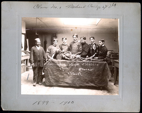
Check it out here. More about this most wonderful of books here and here. The image above is from the book and featured in the Discover Gallery. Caption reads: "Given the outdoor location of this "dissection," along with the students' unusually formal garb, we can be sure this procedure was staged by these students at the U of North Carolina Medical Dept., around 1890. The Seated man is the janitor; the overturned bucket he's sitting on was usually kept at the foot of the dissection table, and was used to collect waste."

Sunday, August 9, 2009

Wellcome Library: 16th C. Anatomical Fugitive Sheets, With Animations Demonstrating Functionality


The amazing Wellcome Library (which I finally had the opportunity to visit this trip to London; more on that soon) has just made their collection of 16th Century anatomical fugitive sheets available on the web. Best of all, the online versions mimic the functionality of the originals; still images are available of the prints at each level of "dissection" (as seen above); you can also watch the prints self-dissect in short, time-lapse films, demonstrating the raising and and lowering of flaps with nary a human hand in sight.

More on anatomical fugitive sheets and their history, in the words of the Wellome Libary Blog:
[Anatomical Fugitive Sheets] depict the human body through labelled illustrations, often using a three-dimensional 'pop-up' device of superimposed flaps, which can be raised in sequence to display the internal anatomy of the male or female figure. The fugitive sheet thus mimics the act of dissection. They were a popular instructional aid in the 16th century and many were produced in vernacular languages which could be read by a lay audience interested in the workings of the human body.
To read the full original post and learn more about anatomical fugitive sheets, click here. You can watch the videos by clicking here. You can view the still images by clicking here and typing in "anatomical fugitive sheet."

Image, from Wellcome Images:
L0017547 Credit: Wellcome Library, London
Anatomical fugitive sheet; female.
Hand-coloured woodcut, Anatomie tresutile pour congnoistre les parties interieures de la femme
Published: Alain de MatonniereParis circa 1560
Collection: Rare Books
Library reference no.: EPB 292/9

Wednesday, June 3, 2009

"Dissection: Photographs of a Rite of Passage in American Medicine 1880–1930" on Science Friday!


If you have not done so already, I highly advise giving a listen to last Friday's episode of NPR's Science Friday. Entitled "'Dissection' Documents Med School Rite-Of-Passage," the clip features a really fascinating interview with the Dittrick Museum's James Edmonson talking about the book Dissection: Photographs of a Rite of Passage in American Medicine 1880–1930 which he recently co-authored with John Warner for Blast Books.

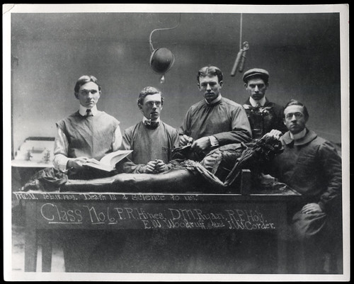
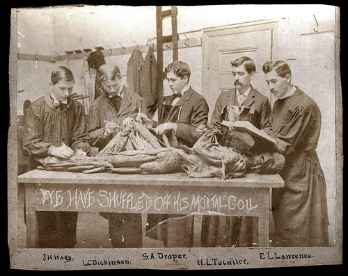
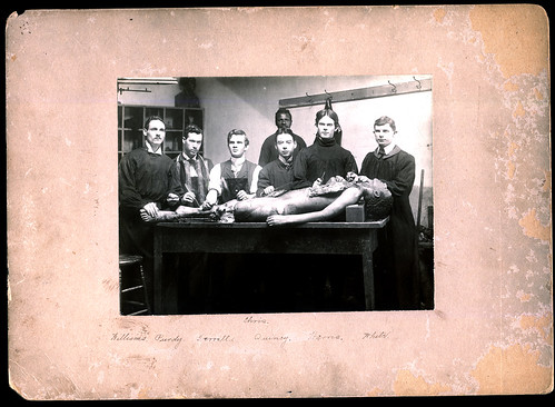
Click here to listen to the interview. More about the book in this previous post. You can purchase a copy of the book (highly recommended!) by clicking here. Images above (click to see larger versions) are drawn from the book, compliments of Blast Books' Laura Lindgren (thanks, Laura!).

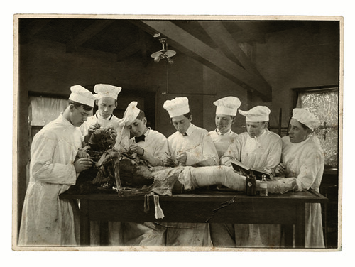
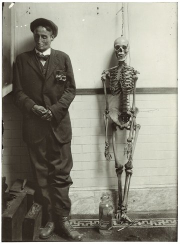
Images: 1) School unknown, ca. 1915. The school and dissectors are unidentified, but in other photographs known to be from the University of Minnesota students also gather around the table wearing distinctive toques. DMHC 2) School unknown, undated. Cadaver, dressed in clothing, stands next to a skeleton, with a fluid-preserved foetal specimen

Tuesday, April 28, 2009

"Dissection: Photographs of a Rite of Passage in American Medicine 1880–1930," Blast Books, 2009


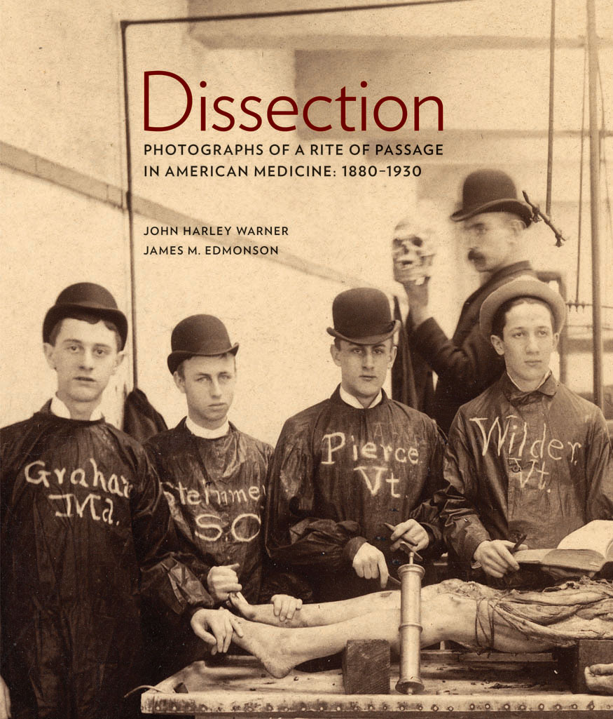
I just got my hands on the new Blast Books offering Dissection: Photographs of a Rite of Passage in American Medicine 1880–1930, by John Harley Warner and James M. Edmonson. You might be familiar with Blast Books from other wonderful publications such as the Mütter Museum calendars and books, and this book is just as memorable and essential as those previous releases. The book is large-scale, beautifully produced, and filled with fascinating and haunting photographs (such as those shown above; click on images see larger versions) so wonderfully reproduced that you feel you could walk right into them. The introductory essays by Warner and Edmonson do an admirable job of parsing this overlooked genre of photography and providing a sense of context for images so unusual and sensational to the modern eye.

I asked Laura Lindgren, co-publisher of Blast Books and the editor and designer of this tome, to give me some information about the book and send me a few of her favorite images (which I have featured above). Here's the press release she sent:
DISSECTION Photographs of a Rite of Passage in American Medicine 1880–1930
by John Harley Warner and James M. Edmonson
Blast Books, 2009

Featuring 138 rare, historic photographs, Dissection is a “landmark book” (Ruth Richardson) that reveals a startling piece of American history, the rite of passage into the mysteries of medicine captured in photography.

From the advent of photography in the 19th century and into the 20th century, medical students, often in secrecy, took photographs of themselves with the cadavers that they dissected: their first patients. The photographs were made in a variety of forms, from proud class portraits to staged dark-humor scenes, from personal documentation to images reproduced on postcards sent in the mail. Poignant, strange, disturbing, and humorous, they are all compelling.

These photographs were made at a time when Victorian societal taboos against intimate knowledge of the human body were uneasily set aside for medical students in pursuit of knowledge that could be gained only in the dissecting room. Dissection, writes Mary Roach, “documents—in archival photographs and informed, approachable prose—a heretofore almost entirely unknown genre, the dissection photograph.”

“Without looking,” writes John Harley Warner, “we cannot see an uncomfortable past and begin to understand the legacies that American doctors and patients live with today.” That uncomfortable past saw the gradual passing of state laws, from 1831 to 1947, to govern the awkward business of cadaver supply—ever inadequate—bringing an end to reliance on professional “resurrectionists,” grave robbing, and dissection as an extended punishment for murder and as a consequence of poverty.

As James Edmonson notes, “Unsettling though these images may be, they are a thread connecting us to the shared experience among medical professionals over generations. . . . As medical schools explore alternatives to human dissection, this rite of passage may disappear.”

Together, the remarkable archival photographs and illuminating essays in Dissection present the astonishing social realities of the pursuit of medical knowledge in 19th- and early-20th-century America.

About the authors: John Harley Warner, PhD, is an historian who focuses chiefly on American medicine and science. In 1986 he joined the Yale faculty with a primary appointment in the School of Medicine, where he is now Avalon Chair of the Section of the History of Medicine. James M. Edmonson, PhD, is Chief Curator of the Dittrick Medical History Center and Museum of Case Western Reserve University in Cleveland.

About Blast Books: Celebrating our 20th anniversary in 2009, Blast publishes books on cultural and historical phenomenons, from Mütter Museum to Mental Hygiene. Visit www.blastbooks.com.

Praise for Dissection
“An extraordinary collection of photographs that makes even today’s flesh-and-blood anatomy laboratories look tame. . . . Forget the truckloads of grandiose prose that has been spun about the art and science of medicine over the centuries: one look at this picture [page 188] and you understand what it is all supposed to be about.”—Abigail Zuger, MD, The New York Times

“This is the most extraordinary book I have ever seen [and] the perfect coffee table book for all the households where I’d most like to be invited for coffee.”
—Mary Roach, author of Stiff and Bonk

“A truly unique and important book [that] documents a period in medical education in a way that is matched by no other existing contribution.”
—Sherwin Nuland, MD, author of How We Die

“What a spectacular book! Warner and Edmonson have amassed an extraordinary array of old dissecting-room photographs. Together, the authors have created a landmark book in the history of dissection in America.”
—Ruth Richardson, D.Phil., author of Death, Dissection and the Destitute and The Making of Mr. Gray’s Anatomy
The book has been (rightfully) been getting a lot of excellent press; see the New York Times review by clicking here and the Slate.com writeup by clicking here; You can also read Bioephemera's thoughtful response here. It is nice to see that audiences are responding so positively and with great interest to what, I was afraid, might be seen as rather off-putting images to the larger public.

Also, I just got back from the MeMa (Medical Museum Association) meeting at the Dittrick Medical History Center in Cleveland, where, after giving a presentation on how medical museums can utilize the web to reach new audiences, I was fortunate enough to see some of the original photographs on display in an exhibition called "Haunting Images: Photography, Dissection, and Medical Students." The Dittrick has created an online version of the exhibition which you can access by clicking here.

I simply cannot recommend this book highly enough. It is absorbing, transporting, beautiful, and thought-provoking. I have never seen another collection like it. You can order a copy of the book (at the moment only about $32 with amazon's discount!) by clicking here. You can see Blast Books other medically oriented illustrated books by clicking here.

Images, top to bottom (after cover):
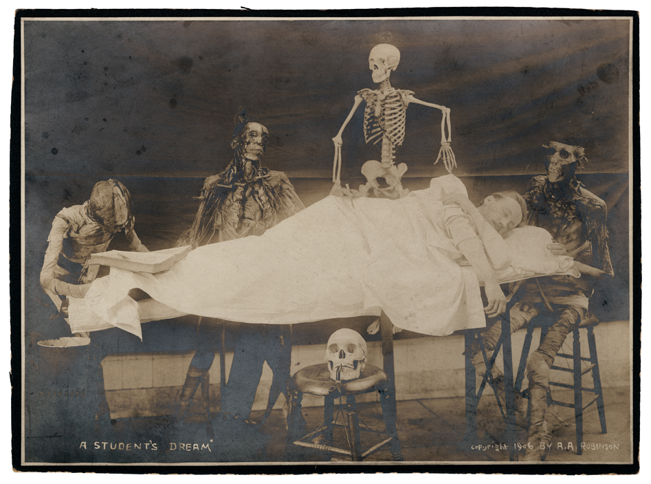
1. “A Student’s Dream,” 1906. Copyright marked A. A. Robinson. A standing skeleton joins seated cadavers, preparing to dissect a sleeping medical student. Iconographic elements that by 1906 had become common-place in dissecting room group portraiture are gathered together in this scene: the book propped open on the “subject’s” feet, the skull resting on a stool in front of the table, and a pipe fitted between the skull’s teeth. DMHC. Used by permission of Blast Books, Inc.

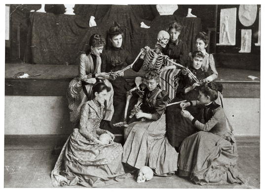
2. Western Female Seminary (renamed the Western College for Women in 1904), Oxford, Ohio. Once removed from dissection, the students at this daughter school of Mount Holyoke studied hygiene, anatomy, and physiology with the use of a skeleton, manikin, and the plaster casts that appear behind the tableau. The instructor probably was Robert L. Rea, M.D., who was resident at the women’s college and later became professor of anatomy at Rush Medical College in Chicago. Miami University Libraries, Western College Memorial Archives, Miami University, Oxford, Ohio. Used by permission of Blast Books, Inc.

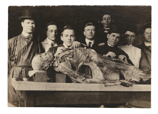
3. Probably Rush Medical College, Chicago, ca. 1915. Private collection. Used by permission of Blast Books, Inc.

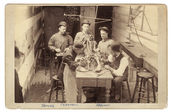
4. Cabinet card dated February 9, 1887, Willamette University Medical Department, Portland, Oregon. A man (wearing a bowler hat, like the dissectors) looks on from the lower left of this cramped space. The medical school had just moved from Salem, Oregon, to Portland. The photograph belonged to Otis D. Butler, age twenty-five when the photograph was taken, who died in 1926 as a result of an infection of the hand received during a surgical operation. DMHC. Used by permission of Blast Books, Inc.

Tuesday, December 16, 2008

"The Anatomy Lesson: Professor Cupid and his Young Pupils," 1918


I have no information on this intriguing and slightly troubling image. I found it on the wonderful Mary Evans Picture Library website, with a caption that merely reads "'The Anatomy Lesson': Professor Cupid and his Young Pupils," 1918."

If anyone has any information about this image, please let me know here.

Saturday, June 7, 2008

Happy Birthday Morbid Anatomy!


This blog's first post was exactly a year ago today. So hey! Happy Birthday to Morbid Anatomy!

Image (from The National Library of Medicine's very excellent Dream Anatomy exhibit: The Anatomy of the Human Gravid Uterus, William Hunter (1718-1783) [anatomist] and Jan van Riemsdyk (fl. 1750-1788) [artist].

Tuesday, June 3, 2008

Tompkins-McCaw Library for the Health Sciences Special Collections, on Flickr!


I just learned, via the blog Bottled Monsters, about the addition of another library collection to the Flickr web-o-sphere: the Special Collections of the Tompkins-McCaw Library for the Health Sciences, Virginia Commonwealth University. Above are some of my favorites, including a collection of students posing with their dissections, but this is just a small taste of what they've got to offer. See the full collection here.

Tuesday, April 1, 2008

Depiction of Human Dissection, 1363


Doing an at-work stock art search today (on a totally unrelated topic) I came across the wonderful image above. Caption reads: "French Manuscript Illumination of a Public Dissection by the Faculty of Montpellier From Guy de Chauliac's Chirurgia Magna, 1363." Chauliac was, apparently, a renowned surgeon of the Middle Ages, with a patient roster inluding a string of Popes.

Tuesday, March 25, 2008

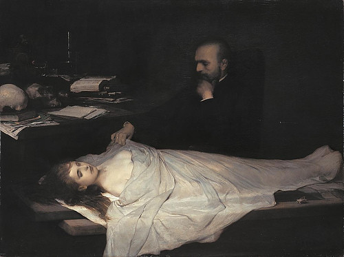
"The Anatomist (Der Anatom)," Gabriel von Max, 1869


This painting brings to mind Edgar Allen Poe's memorable quote: "the death of a beautiful woman is unquestionably the most poetical topic in the world..." It seems this maxim could also--at least circa 1869-- be applied to a beautiful dead woman about to be dissected by a pensive anatomist.

Learn more about the painter, Gabriel von Max, (who studied, among other things, parapsychology, somnambulism, hypnotism, spiritism, Darwinism, asiatic philosophy, and the ideas of Schopenhauer) here. See more images of his work here and here.

Thanks to Ludmilla Jordanova, author of one of my new favorite (and recently cited) books Sexual Visions: Images of Gender in Science and Medicine Between the Eighteenth and Twentieth Centuries,for bringing this painting to my attention. Check out the book for her compelling reading of this and other dissection paintings of the 19th Century featuring beautiful cadavers.

If this topic is of interest to you, you might want to check out a recent Morbid Anatomy post about a dissection print entitled Une Fin A l’Ecole Pratique.