Wednesday, February 27, 2008
Bassett Stereoscopic Dissection Collection (1962)
A small sampling of Stanford University School of Medicine's renowned "Bassett Collection" has been uploaded to Flickr! I really love this new direction Flickr has been taking, working with museums to showcase their collections. (For more on this, see recent posts on Library of Congress and National Museum of Health and Medicine.)
The "Bassett Collection" is more, however, than just a group of beautiful dissection photos; it is also a View-Master set! No joke. This collection premiered in 1962 as the Stereoscopic Atlas of Human Anatomy. This set consisted of 221 View-Master reels of 1,554 color, stereoscopic dissection photos, along with an informational booklet explaining the images. The collection, a collaboration between dissectionist David L. Bassett and View-Master inventor William Gruber, was popular right from the start; when Bassett first exhibited the images, there were lines around the block of people waiting to see them.
This month, they will be released in a new format altogether--online, and with highlighted labeling and audio narration, presumably for a fee. You can view a sampling of the images, fee-free, on Flickr.
Read more about the collection and its history (and future) here. View the Flickr page here. Images of Atlas above from a recent, edited re-release. Thanks to Mike Sappol for sending this along!
Monday, February 25, 2008
Anonymous [Treatise on physiognomy. (Netherlands?, ca. 1790)].
Found on the National Library of Medicine's "Historical Anatomies on the Web." Description reads:
Physiognomy is the science of relating an individual's character, personality, and temperament to the shape of his or her face, head, and/or body. The theories behind it go back to Hippocrates, who believed that physical characteristics of the human body revealed personality traits; Aristotle performed studies on how hair, limbs and facial features predicted personality and temperament. Such theories thrived throughout the Middle Ages and the Renaissance, and the noted Girolamo Cardano (1501-1576) was one of its main proponents. By the 18th century, the study of physiognomy was still taken very seriously as a medical topic, with important additions to the field made by Johann Caspar Lavater (1741-1801). Franz Josef Gall (1758-1828) attempted to make its study even more scientific by measuring human and animal craniums to find correlations between skull shape and behavior, founding the field of phrenology.
The author of this fine, manuscript treatise and sketchbook on physiognomy is unknown. The text is written in Dutch and was probably composed in the 1790s; it is possible that it was created as a dissertation by a medical student.
Labels:
art,
books,
collections,
medicine,
museums,
physiognomy
Saturday, February 23, 2008
Jacques Gamelin (1738-1803)
All images from Nouveau receuil d'ostéologie et de myologie dessiné après nature … pour l'utilité des sciences et des arts … as displayed on the National Library of Medicine's wonderful online exhibit "Historical Anatomies on the Web."
Thursday, February 21, 2008
Extraordinary Bodies: Mütter Museum Photographs Exhibition Opening at The Mütter Museum! Next Friday!
The photographic exhibition Extraordinary Bodies: Mütter Museum Photographs, curated by Laura Lindgren, will soon be appearing at the museum whose collection it celebrates. Originally curated to coincide with the publication of the book The Mütter Museum, this exhibition has traveled nationally since 2002, and will now be presented for the first time at the Mütter Museum. The opening party will be held next Friday, February 29th, from 6-9 PM at the Museum.
The exhibition features images that might look familiar if you were a fan of the legendary Mütter Museum Calendars. Featured photographers include Max Aguilera-Hellweg, Rosamond Purcell, William Wegman, and Joel-Peter Witkin. Their work will be supplemented by reproductions of historic photographs from the Mütter Museum's collection. Click on the invitation above for details.
More exciting (and not wholly unrelated) news: Lindgren plans to revive the Mütter Museum Calendar beginning with 2009; more on that as details become available.
Hope to see you at the opening!
Wednesday, February 20, 2008
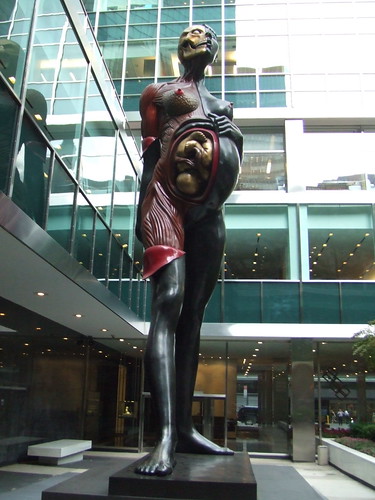
Anatomical Mary, Damien Hirst
Another one from my friend Marie! Damien Hirst's "The Virgin Mother." Displayed in the courtyard of The Lever House, 390 Park Avenue (E 54th St), in midtown Manhattan. Full link here.
Tuesday, February 19, 2008
Reanimation Library Image Archive Launch!
I have been a longtime fan of Andrew Beccone and his small, idiosyncratic "Reanimation Library," which I learned about when it (and he, the helpful and friendly librarian) was featured in the Proteus Gowanus Library/Archives installation. Today he launched his Reanimation Library browsable image archive; as a fan of all things Image Archive, all things Library, and all things Small Idiosyncratic Collection, you will not be surprised to learn that I am pretty excited about this development.
Find out more about the Reanimation Library here; Browse the image archive (from which the above images were drawn) here.
Labels:
image archive,
library
Monday, February 18, 2008
Bill Hayes Reading from his Book The Anatomist, NYC!
I recently finished reading The Anatomist: A True Story of Gray's Anatomy. The book is a curious mix--as much a personal memoir as a biography, and much more of a biography of Henry Vandyke Carter, the illustrator of Gray's Anatomy , then of Henry Grey, about whom very little (apparently) is known.
In the process of researching his subjects, Hayes touches (conversationally and engagingly) on under-known facts of medical history, the world of contemporary medicine and dissection, the imaginative leaps of the biographer, and the drive of a very personal curiosity. I expected not to like the book--I think of the illustrative style of Gray's Anatomy as signaling the shift to the aloof, academic (read: boring and soulless) style of anatomical depiction; I was surprised by how much I did enjoy this book, and am pleased that the public at large seems interested as well.
Tomorrow--Tuesday February 19th--Hayes will read from The Anatomist at 192 Books at 7:00 PM. Find out more info here.
All images from Bartleby's online 1918 version of Gray's Anatomy. View all the illustrations from this edition here.
Friday, February 15, 2008
Another Book for the Wishlist!
My Friend Marie just alerted me to the soon-to-be-published Ephemeral Bodies: Wax Sculpture and the Human Figure. Co-authored by Georges Didi-Huberman, author of another of my favorite (though very theory-heavy) books Invention of Hysteria: Charcot and the Photographic Iconography of the Salpetriere. This promises to be a good one; I seriously cannot wait (and have, in fact, already pre-ordered two copies!)
Thursday, February 14, 2008
More on Anatomical Waxes...From an Unlikely Source
Bonnie Ruberg, authoress of The Village Voice's web column "Heroine Sheik: Sex, Gender, Tech, Culture, and Video Games," sent me this link to her musings on Florentine anatomical waxes. Following is my favorite quote, but check out the whole piece here.
The thing that really fascinates me about these figures, of course, isn’t their creepiness; it’s how they capture a set of historical ideas about sex, gender, and the female body in the form of “science.” It makes you wonder about scientific technology we have today. How is it biased? How does it reveal our own assumptions about sex and gender in between the lines of “fact”? And will it look quite so grotesque to museum-goers in another two or three hundred years?
Photo: 18th century Florentine obstetrical wax held in the Josephinum in Vienna.
Tuesday, February 12, 2008
More Japanese Anatomical Toys!
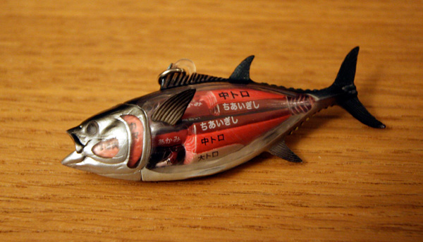
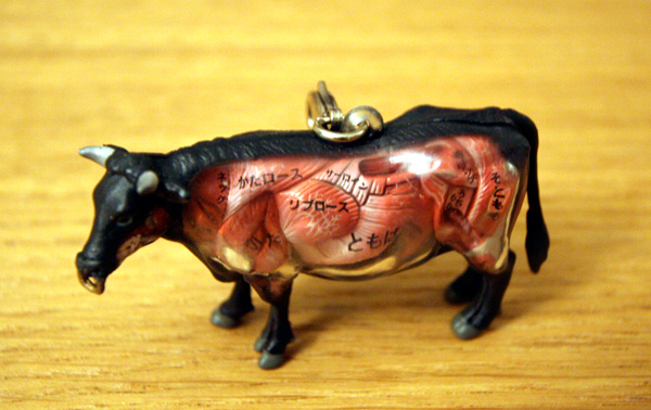
Via Ektopia:Gotochi Dissection Animals--miniature animal models with edible anatomical details called out (see convenient chart on back). Also available: Monkeys and Bears. See Giant Robot for purchasing.
See recent post for more anatomical toys from Japan.
Sunday, February 10, 2008
Wednesday, February 6, 2008
Mysterious Panoptikon Document
In a recent google search, I randomly came across a PDF document called "Panoptikon," filled with intriguing images (such as those above, drawn from the document) and, sadly, many German words I did not understand. I put my Germanaphile friend Stephen Vesecky on the job of translating it, and this is what he came up with:
The first chapter focuses on the psychological appeal of these curiosity shows in Germany during the 19th century. In an era of Victorian morals, it was important to make everything seem scientific, so that it wasn't viewed as bloodlust, to cover up the macabre fascination. As the 1800s wore on, the panoptika were presented more and more as medical museums, but continued to cater to the public's interest in the macabre, as well as the spiritual implications of being able to preserve real human remains and/or create lifelike wax figures, as well as sometimes providing pornographic content. Basically, the Panoptika had a transgressive quality. The book differentiates between some impresarios who presented the show as being of medical or scientific interest, and others who presented it as entertainment. However, there was no doubt that these "curiosity shows" gave important insights into the hidden urges of the bourgeois society of the time.
The Panoptikum offered an incredible variety of exhibits. The text gives some interesting descriptions, which included the sword of Frederick the Great, an enbalmed head of a cannibal from New Zealand, and the journal of a famous sex offender. These items were often faked.
"Oriental Harem," "Witch Torture of the Middle Ages," and "A knothole in the fence around the bathing pool" were some choice titles. It describes the psychology behind the public's interest in these "museums." In those days people were in much closer contact with the gruesomeness of life. More people believed in heaven and hell, and there was a different type of fascination with decay and death. In some ways it catered to the same type of fascination that we satisfy today by watching horror movies. There were "crime galleries" where they had wax figures of well known murderers and would include the tools they used to commit their crimes.
For me, the most amazing thing about the panoptikum is their incredible array of things that were on display, including wax figures of kings and mass murderers, enbalmed sharks, reconstructed torture chambers, and god knows what else.
I urge you to download the PDF for yourself here. If anyone has any further translational comments, or information abou this document or the author, please let me know!
ADDENDUM: I just learned of the identity of the author and the text from where this chapter was drawn--I received an email from Stefan Nagel with a link to his full work "Schaubuden: Geschichte und Erscheinungsformen." I wish I understood German, as this looks like it might be the best book ever... see what I mean by visiting the link. Why is all good research on these topics in French or German?
Tuesday, February 5, 2008
Bones Issue of Cabinet Magazine, 2008
So what went on in the anatomy halls? What did medical students do? They played. With the dead... --"Bone Play," Mike Sappol and Eva Ahren
I have just received my copy of the Bones Issue of Cabinet Magazine. Highlights thus far include Mike Sappol and Eva Ahren's "Bone Play" (from which the above images and quotation are drawn) and Scott F. Gilbert and Ziony Zevit's "Congenital Human Baculum Deficiency," which will make you rethink the biblical story of Adam's rib in the most fascinating of ways. Other articles touch on the mummies of Palermo, paleontology, and trepannation. Well worth checking out.
Note on images: Second image down, caption reads "Photograph from the dissection hall at the Academy of Surgery, Copenhagen, C. 1910; Bottom image, caption reads: A lithograph by Edward Hull depicting Death interrupting an author before his writing is complete, 1827.
The History of Medical Imaging, Warwick University, July 7-11
Read about a wonderful looking upcoming conference on one of my favorite websites: Biomedicine on Display. From the post:
Participants will trace technological developments and their consequences in medicine, alongside consideration of how these new ways of ‘seeing’ the human body reflected and were shaped by the concerns of scientists, physicians, artists, and the general population. The aim of the Summer School is to bring together current and recently completed postgraduates from the humanities and sciences with experts from a number of different fields to engage with a range of technologies for making scientific images of the human body, including the fine arts, drawing and painting, as well as film, photography, X-ray and the current medical imaging techniques of digital biomedicine... What are the epistemological, moral and philosophical consequences of our desire to picture all functions of the human body? What does it mean to be human in a world of global mass media in which the individual body is central, yet increasingly public and commercialised? Are there alternatives to the understanding in Western science since the nineteenth century that vision is the primary avenue to knowledge and sight takes precedence over the other senses as a tool in the analysis of living things?
Check out the whole post here. See the full program here.
Friday, February 1, 2008
Mortality, Radio Lab, 2007
I just heard the most fascinating story on NPR's Radio Lab--the program is called "Mortality" and originally aired in June of last year. Via interviews with scientists, researchers, and artists, it examines mortality from the cellular to the societal level; in the process, it touches on such curiosities as "the grim reaper cell," "The Hayflick Limit," and the Japanese tradition of "throwing grandma away."
Listen to the story (and read more about it) here. HIGHLY recommended.
Image: Death and Life, Gustav Klimt, 1916
Subscribe to:
Posts (Atom)