Monday, April 28, 2008

"Anatomical Theatre" Website Launch!


Finally, after many months, I am launching a somewhat finalized draft of the Anatomical Theatre website. Herein you will find an a expanded version of the physical exhibition, which features 64 photographs (the website features nearly 100!) of medical museum artifacts held in great collections in Europe and the United States. The artifacts range from preserved human remains to models made from ivory, wax, and papier mâché, with provenances spanning from the 16th to the 20th centuries. As much information as possible is coupled with each image. Visit the website to learn more about this project, find information about featured museums, and view the photographs.

Anatomical Theatre: Depictions of the Body, Disease, and Death in Medical Museums of the Western World is a travelling exhibition; next location to be announced.

Note: I attempted to include as much factual data as possible about the artifacts photographed. Often, I was the only one checking facts. if anyone comes across any mistakes please drop me a line and let me know.

All images above from the Anatomical Theatre website; visit the website to find out more information about each piece.

Friday, April 25, 2008

Padre Pio's Corpse Exhumed, Displayed in Glass Coffin after 40 Years in the Grave. Really.


Its not everyday that one gets news of an exhumed corpse being exhibited to hordes of tourists in a glass coffin; today, just such news came my way.

Yesterday, the corpse of Padre Pio was put on display in an elaborate glass-and-marble coffin after his body was removed from the vault in which had resided for 40 (!!!) years after his death at 81 years of age. His body was too far decayed, it is reported, to be exhibited without enhancement; his head is covered with a life-like silicon mask prepared by the company that supplies figures for Madame Tussauds; his true fingers are visible, blackened and half-hidden by half gloves. Reportedly, his hands do not bear the stigmata they often did in life.

He was exhumed, reports The Independent, "so the condition of the body could be ascertained before being consigned to its permanent home in a crypt under the town's vast modern church." (The report continues: "Officials who examined the corpse said it was in 'fair condition', apart from the head, much of which had been reduced to bare bone. A team of medical scientists and biochemists has been working since then to restore the corpse to a presentable condition.")

Padre Pio was an interesting and complex figure; In his time, he had a cult-like following that revered him as a saint, while others accused him of being a fake, even going so far as to accuse him of using carbolic acid to create the stigmata he famously displayed on his hands and feet. Pope John Paul II canonized him into sainthood in 2002.

The Los Angeles Times has a great article on this, from which this video (and much of the information in this post) is drawn; read it here.

Yet MORE on Japanese Anatomical Charts!


Yes, yet MORE on the Japanese Anatomical Charts discussed in two recent posts (here and here). Now we have a translation! The folks at Pink Tentacle have written a full story on these charts, complete with names, dates, and history. They have even thrown in some newly reassembled images (see above).

We discover that these are called the Kaibo Zonshinzu Anatomy Scrolls (1819) and were painted by Kyoto-area physician Yasukazu Minagaki (1784-1825) under the tutelage of Dutch anatomist Philip von Siebold, the first European to teach Western medicine in Japan. We also learn that they are regarded as the finest collection of Japanese early 19th-century anatomical drawings, and that the subjects dissected and portrayed were "heinous criminals executed by decapitation."

So pleased that a translator was able to translate this page and answer so many of my questions. Check out the full story here. And see the original collection here.

Tuesday, April 22, 2008

Japanese Anatomical Charts Expanded


The fine folks at Ectoplasmosis have taken the trouble of assembling some of those Japanese anatomical charts I posted about yesterday into their intended scroll-like continuum. Not as surreal, but lovely none-the-less. Check out their some of their handiwork above. See their post here.

The Bassett Stereoscopic Dissection Collection in the New York Times!


In the science section of today's New York Times, there is an article about the amazing Bassett Stereoscopic Dissection Collection (as discussed in greater detail in a previous Morbid Anatomy Post.) The project was a collaboration between William Gruber, the inventor of the View-Master, and Dr. David L. Bassett of Stanford University. The product: the 25-volume “Stereoscopic Atlas of Human Anatomy” released in 1962 consisting of thousands of images illustrating human anatomy (see above) on hundreds of View-Master reels.

The article touches on the history of the project, the partnership between Bassett and Gruber, and how the collection was received when it was released. It goes on to detail Stanford University's plans to digitize the collection and charge access to it; those with 3d glasses will be able to view them on their computer in their original 3-dimensional glory.

Check out the full article here; check out the slide show here. And click here to see the Flickr page launched by Stanford to showcase the images.

Thanks so much to Pam of Phantasmaphile for sending this my way.

Monday, April 21, 2008

Japanese Anatomical Charts


Thanks to Peacay of Bibliodyssey for sending me the link to these surreally fascinating (and mysterious, as I understand no Japanese) Japanese Anatomical Charts. You can peruse them all (this is just a small sampling) here.

Friday, April 18, 2008

Translation of Mysterious "Panoptikon" Document


Thanks to Sebastian Keller for kindly translating the Mysterious Panoptikon Document." There is much great information to be found here, and a wonderful collection of quotations. Here is a sample:

“I entered a display of waxworks; the entourage of the ruler looked very slutty and neglected. It was a terrible loneliness and I hastened through to a closed room with the collection of anatomy on display. There almost any part of the human body was to be found on display, made of wax, most of them in sick, terrible states, a really strange assembly of human conditions. A big part of that assembly was constituted of a long row of glasses which contained all the stages of a foetuses growth. Those weren't wax, but real beings and they were sitting in the alcohol like philosophers. Their thoughtfulness was all the more clear as those guys should have been the youth of that assembly. But suddenly in the hut right next to that there was loud music and drums an the wall shuddered and all the quiet attention vanished. Those little beings started to shiver and dance in a wild polca and soon there was anarchy, so I don't believe that this assembly ended with an address.” (Gottfried Keller, dream diary, S.93f)...


Read the entire translation (and add your own comments) here; compare it against Stefan Nagel's orginal "Schaubuden - Geschichte und Erscheinungsformen" here. You can find additional images here.

Pictured above: Page 24 from chapter 1 "Panoptikum, Kuriosa" of l "Schaubuden - Geschichte und Erscheinungsformen."

Wednesday, April 16, 2008

Florentine Anatomical Venus in Rare American Appearance!


The Getty Villa in Malibu, California has an interesting show up called "The Color of Life: Polychromy in Sculpture from Antiquity to the Present." The exhibition showcases the many ways artists have used color in figural sculpture for centuries, and, to my excitement, includes an actual Anatomical Venus from the famed La Specola collection in Florence!

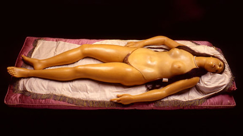
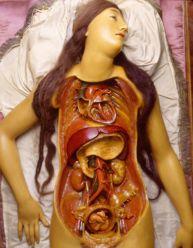
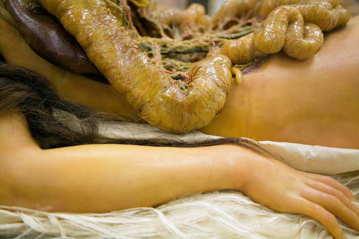
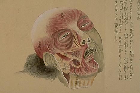
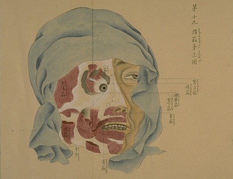
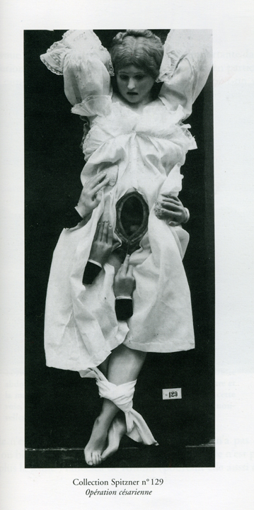
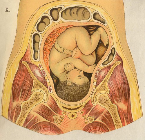
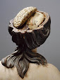
Anatomical Venuses are life-sized wax anatomical models of idealized women, extremely realistic in appearance and often adorned with real hair and ornamental jewelry. These figures consist of removable parts that can be "dissected" to demonstrate anatomy-- a breast plate is lifted to reveal the inner workings of the mysterious female body, often with a fetus to be found nestling in the womb (see before and after above). This was a way to share anatomical discovery with a larger audience without the need for an actual human dissection.

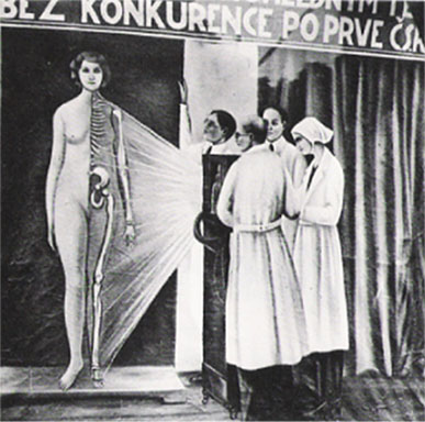
Anatomical Venuses are probably the most historically popular form of anatomical models; in the 19th-Century, they were the centerpiece of museums and itinerant shows of all kinds, and possessed great power to draw crowds. The 18th-Century Florentine Venuses are the best remembered today, in no small part due to Taschen's Encyclopaedia Anatomica,and are considered, by some, to be the finest examples of Anatomical Venuses known to exist.

This Anatomical Venus featured in the Getty show, completed in 1782 by Clemente Susini and his workshop, is truly a masterwork of the genre, and outshines the many copies held by medical museums throughout Europe who, impressed by the veracity and workmanship of the Florentine Venuses, commissioned their own from Susini's workshop. For example, the core of the collection of the Viennese Josephinum Museum consists of 1192 models commissioned by Emperor Joseph from the Susini workshop in 1784 for the training of his military doctors. As a body of work they are interesting, but somehow the models in this collection pale in comparison to the La Specola models. The workmanship is a bit shoddier, the visages a bit less alluring.

The Anatomical Venus exhibited in the Getty exhibition is one of the finest, and almost never leaves her home in Florence--she has only been transported twice in her long history, and even has a specially-built traveling case to protect her delicate wax body. This means that, sadly, you will not see her in her original setting--an elegant rosewood and Venetian glass case-- but the relative accessibility of the piece (i.e. in the United States) should make up for this lack.

It is really great to see anatomical models being seriously approached as artwork in an exhibition of this sort; I consider it a bold move on the part of the curators, and cannot help wondering what strings the curators had to pull in order to acquire one of these rare and fragile Venuses on loan. It is also nice to see that, in reviews of the show, the Venus seems to be a real standout. This supports my belief that, if more people knew of these Venuses and other anatomical models, they would be seen as intriguing artworks and cultural documents, worthy of a greater amount of study as well as inclusion in the medical art canon.

From the UCLA paper The Daily Bruin:
The two most impressive pieces of the whole exhibition, which may please even marble lovers, both shock and fascinate at the same time. One of them is an 18th-century wax model of a nude life-size woman called the “Anatomical Venus,” which shows the multicolored exterior and interior of a female body with an almost uncanny precision.

And from the LA Times review of the show:
The strangest, without a doubt, is an 18th century wax figure known as the "Anatomical Venus": a comely young woman, life-sized and nude, lying prostrate on a pink silk cushion in what looks to be a state of sensual rapture, her torso flayed and all her glistening organs -- including a womb containing a tiny fetus -- revealed. Her long brown hair is real, her eyes are open and unfocused, and the cloth of her pillow is crumpled -- she might as well be writhing. The product of one sculptor's clearly intimate experience with cadavers, she suggests an Enlightenment-era St. Teresa ravished by communion with the invisible forces of science.


To learn more about the show, which runs until June 23 of this year, visit the exhibition website, complete with gallery slideshow. Better yet, visit the show in person if you are able. This might be your only chance EVER to see an Anatomical Venus of this quality without traveling to Florence. To see more images of Anatomical Venuses and other anatomical models, see the Anatomical Theatre Gallery. To learn more about medical models, see Susan Lamb's An Analysis of Anatomy Models and A. W. Bates' abstract for Anatomical Venuses: The Aesthetics of Anatomical Modeling in 18th- and 19th-Century Europe. You can also see the Venus in all states of metaphorical undress in Taschen's Encyclopaedia Anatomica.

Thanks to the Getty for supplying the above images and answering my barrage of questions. Photographs of the Anatomical Venus by Saulo Bambi, Museo di Storia Naturale "La Specola"; Florence, Italy

And if any Morbid Anatomy readers are in the Los Angeles area and could take a photo of the model in context of the show and send me a copy, I'd very much appreciate it.

Tuesday, April 15, 2008

"The Spitzner Museum's Wax Woman" Comicbook!


Morbid Anatomy reader Heather Whiteside alerted me to the existence of a comic book called "The Spitzner Museum's Wax Woman," by Francoise Riviere and Andreas Martens. She found out about it in the 2001 "Taboo" Issue of Heavy Metal Magazine.

I just purchased a copy of the magazine, but I am dying to find a copy of the original comic book. If anyone has any leads on where to find one and could point me in the right direction, I would be most grateful. Further, the kind folks at Kikkerland Design have offered to help me on my quest, by donating an Anatomical Eye 3-D Puzzle to any person who can help me acquire a copy.

Image from Voir - La Collection Spitzner, eds. Phillipe Blon and Stephen Bann, 1998; visit the Morbid Anatomy Library to see more images from the book. For more on the Spitzner waxes, check see recent Morbid Anatomy posts Musée de la Médecine and Surrealism and Anatomical Waxes.

Friday, April 11, 2008

More images from Mysterious German Manuscript


Won't some kind soul please translate Stefan Nagel's online illustrated manuscript "Schaubuden: Geschichte und Erscheinungsformen" for me? The images are so good, I am dying to know the context.

For more on Nagel's manuscript, see this recent post. View the images in context here. See more images from the document in the Morbid Anatomy Library.

Thursday, April 10, 2008

Richard Russell's Anatomical Collage Work, 20th C


I am not usually a big fan of collage, but I find myself drawn to the work of Atlanta based artist Richard Russell, and not just because of the use of anatomical ephemera in his works.

You can see more of his work on his online portfolio and Flickr page.

Thanks to Street Anatomy for bringing Russell's work to my attention.

Tuesday, April 8, 2008

Professor Palmer's "Office" (Medicine Stage Show), c. 1860


Just got the book A Morning's Work: Medical Photographs from the Burns Archive & Collection, 1843-1939 (published by Twin Palms Publishers in the mail yesterday. The book seems to comprise a sample of Dr. Stanley Burns' broad and vast collection of photographs related to death, the body, and medicine. Have not had the time to peruse it properly, but, at a glance, it looks amazing. Here is one of my favorite images thus far.

Saturday, April 5, 2008

Anatomie Modèle Femme (1937)


From the wonderful ephemera collection showcased on the Agence Eureka blog.

Wednesday, April 2, 2008

Anatomical Flipbook, 1939


I have long been a fan of the Agence Eureka blog; yesterday I made the leap to the associated Flickr page, which is a treasure trove of paper ephemera.The above images are from a Flickr set entitled "Anatomie Femme 1939;" check out the complete set here.