Showing posts with label medical museum. Show all posts

Tuesday, April 1, 2014

On the Collection and Display of Human Remains: Guest Post by Evan Michelson, TV's "Oddities" and Morbid Anatomy Museum

The debate about the ethics of collecting and displaying human remains went mainstream recently with this CNN article about a man who stole over sixty human brains and other specimens from the Indiana Medical History Museum and tried to sell them on Ebay. Morbid Anatomy Museum's scholar in residence Evan Michelson is a researcher into the history of such collections, in contexts both sacred and secular. Following is her thoughtful and considered response to the CNN article, which went so far as to single out her show "Oddities" as "being illustrative of a growing trend for collecting curiosities, particularly anatomical specimens."
A CNN article published on January 3, 2014 chronicled the arrest of a young man who stole some early brain specimens from an Indiana medical museum to sell on Ebay. In the article the TV show "Oddities" was cited as being illustrative of a growing trend for collecting curiosities, particularly anatomical specimens. Said the executive director of the museum: "it's definitely bizarre. It's infuriating that they do not have respect for the human remains." This statement raises a few important points: I think everyone can agree that the illegal buying, selling and hoarding of exhumed or pilfered human remains is deeply disrespectful, repugnant, and indefensible on moral and legal grounds. No one can condone or defend such ghoulish goings-on. What is not being addressed, however, is an unavoidable truth: humans have always lived with, loved, and learned from our dead.

The urge to collect, display and venerate human remains is nothing new: it stretches back through the millennia, and plays a vital role in the history of science, medicine and many religions across cultures and around the globe.The widespread practice of ancestor worship originated at a time before recorded history (and is still practiced to this day). The gathering of bones is an irrepressible and primal human urge. Humans have long honored our dead with altars, elevating bones (particularly skulls) to a level of intimate spiritual totem. In many cultures the presence of human remains brings both comfort and continuity. From the Tibetan Kanling (a flute made from a human thigh bone) to the mummies of Palermo to the gorgeous calligraphy of 19th century French memorial hair work, to be in the material presence of the dead is to be one with generations past, to commune with the spirits, to ask favors, to remember, to harness power and to connect with the infinite.

In the service of science and medicine, human remains (such as those pilfered from the museum) have long been essential. It is only through contact with the dead that the secrets of the living have been revealed. The great anatomical insights of the classical physician and philosopher Galen (who primarily studied the anatomy of primates and pigs) are often overshadowed by the many glaring inaccuracies. These fatal mistakes ruled the study of anatomy for more than 1300 years, until anatomists like Andreas Vesalius delved into the human body proper to uncover a more accurate and comprehensive map of our internal architecture. In the 16th century depictions of these anatomical discoveries entered our collective human consciousness, and human dissections became works of high art and an essential part of the great humanist movement that flowed through the Renaissance and powered the scientific revolution. There followed the era of the beautiful corpse, when ceroplasts like Ercole Lelli and Clemente Susini created wax corpses and anatomical moulages of such surpassing beauty and accuracy that they inspired Popes, Emperors and commoners alike to see human anatomy as an important discipline worthy of respect and wonder. The human corpus had at last become a part of high and low common visual culture.

The preservation and display of actual human remains is a time-honored tradition in the great Positivist cities of the Western world, and most centers of learning had their own anatomical collections. These specimens of human anatomy were artfully prepared and displayed, and they illustrate the collective human journey from the realm of superstition through the refinements of natural philosophy and eventually to the rise of modern science. Exhibitions like "Body Worlds" still draw large crowds, eager to examine up-close what is so often kept hidden, and so often considered taboo. The sourcing of the "Body Worlds" cadavers is cause for justified legal and moral scrutiny, but their public display is an enlightening, time-honored tradition. For centuries, museums of anatomy have housed human specimens that are at once didactic, metaphorical and breathtakingly beautiful. These anatomized specimens can still be seen on exhibition in museums and in private collections, and they still provide unparalleled insight into our earthly selves. Anatomy is now digitized, and our bodies (down to a microscopic level) are available at the click of a button, but there is no substitute for the visceral presence of preserved anatomy; it is the best way to know ourselves.

Nowhere is the power of human remains more evident than in the evolution of the Christian religion and the rise of the Roman Catholic Church; there the collection and adoration of human body parts reached its artistic and spiritual pinnacle. The cult of the saints guaranteed that human remains would take center stage in the evolving political, economic and spiritual journey of the West. Religious pilgrims travelled great distances to be in the presence of the bones of the early martyrs, and the wealth thus generated drove an unprecedented competition for relics and a trade in human body parts (particularly in Western Europe) that determined the power centers of the modern world. We are all living in a map shaped by the preservation, display and possession of the dead.

The Temple of the Tooth in Kandy, Sri Lanka is home to the tooth of the Buddha, one of the most celebrated relics on Earth. Once a year the relic is featured at a 10 day festival that includes fire dancers, musicians, street performers and scores of elephants. It draws an estimated crowd of one million participants, making it one of the largest Buddhist gatherings in the world. It is obvious that there is something irresistible about our anatomy, something that reaches us on a primal level. We fear and worship human remains, we shun death but we are irresistibly drawn to the dead. That young man who stole those brains broke the law and showed great disrespect in the commission of that crime. The instinct to collect, display and commune with the dead, however, is not as bizarre or disrespectful as some may think: it connects us with our earthly selves, and allows us to glimpse eternity.
Image sourced here.

Thursday, January 2, 2014

Museum of Mexican Medicine (Museo de la Medicina Mexicana), Palace of the Inquisition, Mexico City

On our recent trip to Mexico, we stopped in at the Museum of Mexican Medicine (Museo de la Medicina Mexicana) located at the Palace of the Inquisition in Mexico City. On the Sunday we went, it was so completely mobbed that we could barely even push our way up through all the families taking cell phone photos of the amazing moulages to see much of them ourselves. It was lovely to see such a museum so popular on any day, let alone a Sunday.

You can see some of the photos from the trip above; click here to see a more complete set. You can find out more about the museum by clicking here.

Monday, December 16, 2013

Call for Papers: ‘Between Medical Collections and Their Audiences’ – EAMHMS Congress, London 2014

This excellent looking call for papers just in from our friend Katie Maggs of the Science Museum! If interested, you can send abstracts to her at katie.maggs [at] sciencemuseum.ac.uk. Submissions must be in by 20th January 2014.

Full details follow:
Call for Papers: ‘Between medical collections and their audiences’ – EAMHMS Congress, London 2014
EAMHMS (European Association of Museums of the History of Medical Sciences ) Congress, London 2014
‘Between medical collections and their audiences’
September 4th – 6th 2014
Science Museum, Royal College of Surgeons of England, Wellcome Collection
The European Association of Museums of the History of Medical Sciences Congress biennial meeting will be held in London, September 4th – 6th 2014, jointly hosted between the Science Museum, the Royal College of Surgeons (Hunterian Museum) & the Wellcome Collection.

The theme for the 2014 Congress is ‘Between medical collections and their audiences’.

After a thrilling conference of the European Association of Museums of the History of Medical Sciences (EAMHMS) hosted by the Charite, Berlin 2012, we would like to invite the members of the association, as well as interested scholars and curators from the community of medical history collections and museums to participate in the next meeting of the organisation – London 2014.

EAMHMS is an active global network of curators, scholars & stakeholders with an interest in medical collections. The biennial Congress is a great opportunity to present research within a vibrant forum of debate and discussion, and promotes international exchange and collaboration amongst medical history museums. The Association, although nominally European, today attracts participation from around the world and is thus the leading international body of medical museums and collections.

The 2014 Congress will focus on medical collections and audiences – looking at how medical heritage is used to intrigue, problematise, teach, and stimulate interaction and reflection about medicine of the past and of today. Who engages with our collections and which new audiences are we innovating ways to engage with? Our visitors are not passive spectators, but rather participants in the creating of and telling of stories about medical heritage – so what does that mean for the future of curating medical collections & displays?

We welcome contributions around strands such as:
  1. Retelling ‘old’ histories with new narratives – in what ways can we reinvigorate audience engagement through re-telling established histories of medicine?
  2. Medical professionals as audience – in what ways can we connect and involve our collections to medical students or retired clinicians and beyond?
  3. Communicating problematic medical heritage – such as medical controversies, human remains, medical ethics.
  4. Audiences and communicating contemporary medical practise – how might we foster contemporary collecting or engage visitors with the material culture of contemporary biomedical practices.  
  5. Histories of audiences visiting medical collections - who has historically visited medical museums, how has this changed, what impact does this have?
  6. Audiences beyond the walls of the medical museum - approaches to engaging audiences in virtual world or through outreach.
  7. Research & innovation with audiences - what do we understand about our visiting audiences through audience evaluation? What new interpretative approaches excite us?
Papers are requested to be no more than 15 minutes in length (with an additional 15 minutes allotted for questions). The language for abstracts, talks, and discussions will be English. Short abstracts will be circulated to Congress participants in advance.

As spaces for speakers are limited, we would also like to invite contributions in the form of poster presentations – which will be discussed in a dedicated session.

We ask you to choose a topic from the above-mentioned issues and send your paper or poster abstract (maximum 1000 characters) with a title, your name and brief biography, the name of your institution (if you are attached to any) and your contact details (preferably e-mail address) to Katie Maggs, Curator of Clinical & Research Medicine at the Science Museum, London:  katie.maggs@sciencemuseum.ac.uk.

Deadline for submission: 20th January 2014.

A programme committee will select abstracts for an inspiring programme. Speakers and poster presenters will receive confirmation by mid-March. If your contribution is chosen, you will be asked to send in an extended abstract (2 to 5 pages) April 2014.

EAMHMS shares costs across the congress, so unfortunately speakers do not go free. The conference fee is envisaged at about £200, but will be confirmed shortly. We look to minimise costs wherever possible.

There may be a reduced fee for a small number of student contributors.

General enrollment for this conference will open in April 2014. We’re looking forward to a thrilling conference! See you in London 2014.
Image: audience interaction at the pathological museum in Mexico City, 2013.

Thursday, June 13, 2013

In Search of Medical Museum Books

On the heels of the publication of the new book Medical Museums: Past, Present and Future, I am working to assemble a master list of all known medical museum books and catalogs. Many of these museums are quite small and obscure, and thier publications, if they exist, hard to find.

If any Morbid Anatomy readers happen to know of any such publications, no matter how humble, I would greatly appreciate if if you could let me know! You can do so by emailing me at morbidanatomy [at] gmail.com.

You can find also out more Medical Museums: Past, Present and Future--with its "17 richly illustrated chapters" covering collections such as Berlin's Charité, the Copenhagen Medical Museion, Edinburgh's Surgeons’ Hall, La Specola of Florence, London's Hunterian and Wellcome Collection, the Mütter of Philadelphia, Morbid Anatomy, and much more!--by clicking here; you can buy a copy of your own by clicking here.

Thursday, May 16, 2013

This Saturday, May 18th: Celebrate The London Hunterian's 200th Birthday With Lectures, Reception, and Book Release Party for "Medical Museums: Past, Present and Future" With Chapter by Morbid Anatomy!

This Saturday, May 18th, London's Hunterian Museum will be celebrating its 200th birthday (!!!!!) with a series of lectures, a late view of the new exhibition, and a reception and party for their new, lavishly illustrated and utterly essential collection of essays showcasing medical museums around the world entitled Medical Museums: Past, Present and Future.

The lectures--which begin at 4:30 PM--and reception and late view--beginning at 6:00 PM--are free but require a reservation; a special dinner beginning at 7:30 PM will run you £65; you can RSVP for all at 020 7869 6568.


The book--whose cover you see above--was edited by Hunterian director Samuel Alberti and Dr. Elizabeth Hallam, Universities of Aberdeen and Oxford to commemorate the museums' bicentennial, and is richly illustrated with hundreds of photos of many of the worlds most wonderful medical museums. You will also notice a wee chapter of my own, which features many never before shared photos of medical museum back rooms.

You can find out more about the book in the following press release, and can order a copy by emailing shop [at] rcseng.ac.uk or calling 020 7869 6562.

Hope very much to see you at Saturday's festivities!

Medical Museums: Past, Present and Future
Edited by Samuel J M M Alberti and Elizabeth Hallam

This book brings together a unique collaboration of curators and scholars from Europe and the United States to open up new perspectives on the past, present and future of medical museums.

Offering readers unrivalled access to international collections of preserved animal and human bodies, instruments and art, Medical Museums features many previously unpublished images and untold stories from this fascinating and disturbing world.
Medical Museums is a bold, eclectic anthology that offers readers a unique opportunity to experience this compelling heritage through clearly written, well-informed text and sumptuous images. Insightful essays informed by current debates in medical history, anthropology, museology and visual studies are complemented by astonishing images provided by both well-known and niche museums from around the world. With unparalleled coverage provided by curators, the 17 richly illustrated chapters explore collections from Aberdeen to Zurich including: Berlin (the Charité), Cleveland (the Dittrick), Copenhagen (the Medical Museion), Edinburgh (Surgeons’ Hall), Florence (La Specola), Leiden, London (the Hunterian Museum, the Science Museum, and Wellcome Collection), Philadelphia (the Mütter), Stockholm (the Karolinska Institute), Washington DC (the National Museum of Health and Medicine and the Smithsonian).

Published by The Royal College of Surgeons of England to commemorate the bicentenary of the Hunterian Museum.

Paperback, 256 pages; H 23, W18cm; 250 b/w and colour illustrations (Code 879)
You can find out more about the events here, and order a copy of the book by emailing shop [at] rcseng.ac.uk or calling 020 7869 6562.

Friday, May 10, 2013

Early 19th Century Corrosion Cast of a Child: Guest Post by Carla Valentine, Curator of Barts Pathology Museum

I am so excited to announce that the amazing Barts Pathology Museum has launched a blog! On the blog, curator Carla Valentine will regularly report on her detailed research into the over 5,000 fascinating specimens she cares for at the Grade II listed Barts Pathology Museum at St Bartholomew’s Hospital, West Smithfield, London.

To celebrate the launch, I asked Carla if she might like to write a guest post about one of my favorite pieces from the collection which I had admired in the recent Museum of London's "Doctors, Dissection and ResurrectionMen" exhibition.

Following is her post; If you like what you read (and see!) I  urge you to check out the wonderful--and delightfully image heavy--blog by clicking here.
After a short stint at the Museum of London's 'Doctors, Dissection and Resurrection Men' this year I’m happy to say the above specimen – a corrosion cast of a child covered in shellac - has found its new home here at Barts Pathology Museum. It was originally housed at our sister hospital The Royal London, which is of course the home of Joseph Merrick (The Elephant Man) and in fact it was Frederick Treves, the man who ‘discovered’ the Elephant Man, who actually purchased this specimen on a trip to Paris in the 1800s.
We don’t have much more information about the child except that it probably pre-dates Treves by a long way and is from the early 1800s.
‘Corrosion casting’ similar to this was developed by such pioneers as Jan Swammerdam (above, Wikicommons) and Frederick Ruysch (below, Wikicommons) as early as the 16th century and was still popular with eighteenth- and nineteenth-century anatomists. Swammerdam and Ruysch had created a technique to inject the blood and lymphatic vessels of corpses with a mixture of wax, talc and pigments that set and endured, and often the surrounding flesh was corroded or removed. Anatomists were still using Swammerdam’s syringe to inject mixtures of varying proportions into their specimens until the early 20th Century.
According to Museum Vrolik, Ruysch had created the art of injecting specimens with a wax-like substance, dyed red with cinnabar, so that they could be more lifelike when displayed in jars (similar to the pink-tinted embalming fluid used to make loved ones more lifelike today.) However, he also injected tissues and organs which he then ‘embalmed and dried’ which would be more likely to give the dehydrated look of this specimen. In addition to such methods Ruysch introduced a new way of embalming specimens (based upon techniques already known by the ancient Egyptians).”
 
The passage reads: “
Examples of organs in the Vrolik collection that have been injected with wax or dried include placentas and penises and, in particular, human and animal hearts. In order to make their anatomical structure more visible, in some cases one half of the heart was injected with red wax and the other half of the organ was injected with dark blue wax. Dry specimens, like bone and certain membranes, were dehydrated by exposing them to the external environment and subsequently coating them with a kind of varnish. The majority of the dry organ and tissue specimens were coated with shellac after drying.” A fate which seems to have befallen our shellac child in his entirety.
Shellac is an incredibly versatile substance which is secreted by the female Laccifer lacca (Lac Beetle), an insect of the order Hemoptera or the ‘true bugs’. It’s well known for it uses as a varnish for garden fences, paintings and musical instruments, but perhaps more surprising is the prevalence of edible shellac compounds in the pharmaceutical and food industries: it is found on medicinal capsules, in the wax of apples and lemons and on confectionery such as M&Ms. Used to limit water loss and prevent dessication, as well as to limit entry of pathogens, it is fairly obvious why this would be a good preservative for prepared anatomical specimens.
The interesting thing about this specimen is that it looks old and of its time – perhaps by using techniques mirroring those of the Ancient Egyptians, Ruysch ensured that an appearance of antiquity was an inevitable by-product. Medical students faced with a specimen such as this today possibly would not value its use as a teaching aid as much as they would have in those heady days of the 18th and 19th Centuries and would relegate it to the realms of ‘interesting’ and ‘unusual’ artwork but nothing more.
However that shouldn’t be the case – a study from 2011 was carried out to illustrate the use of shellac as a modern preservation method to replace the dangerous and carcinogenic formalin/formaldehyde which is currently used in dissection rooms. This study (Ref 2) has shown that shellac will preserve a new cadaver indefinitely in a way that is non-toxic, and said cadaver can also be placed into a softening room and in three days’ time be ready for student to use in their dissections. So it seems that an outdated yet beautiful specimen such as this can be used to inspire future generations of medical students and will also be a fascinating talking point on the ground floor of Barts Pathology Museum.
You can find out more about Barts Pathology Museum by clicking here. To can check out their new blog by clicking here. You can find out more about Carla Valentine by clicking here.

Image credits: Top image: Joanna Ebenstein; all other images of the specimen: Barts Pathology Museum, QMUL; portraits: wikimedia

Monday, March 11, 2013

Museo di Anatomia Umana (Museum of Human Anatomy), Pisa: Italy Trip Guest Post by Evan Michelson, TV's "Oddities" and Morbid Anatomy Library


Below, the fourth guest post by Evan Michelson of "Oddities" and the Morbid Anatomy Library documenting our trip through Italy researching the history of the preservation and display of the human corpse. Here, her response to the small but wonderful Museo di Anatomia Umana (Museum of Human Anatomy) of Pisa:
The Museum of Human Anatomy at Pisa is located at the school of medicine and surgery, just a few steps away from the Piazza dei Miracoli which contains the overly-familiar Leaning Tower - a 12th century campanile gone wrong.

The museum collection has an interesting history: Pisa was host to the First Conference of Italian Scientists in 1839 - a somewhat radical gathering uniting scientists from several disciplines. According to our guide, this was a philosophically Positivist convocation that was determined to make sure that science would have an important role to play in a newly unifying Italy. Scientists from the Papal States were not in attendance. Much of the current museum collection was organized for that gathering.

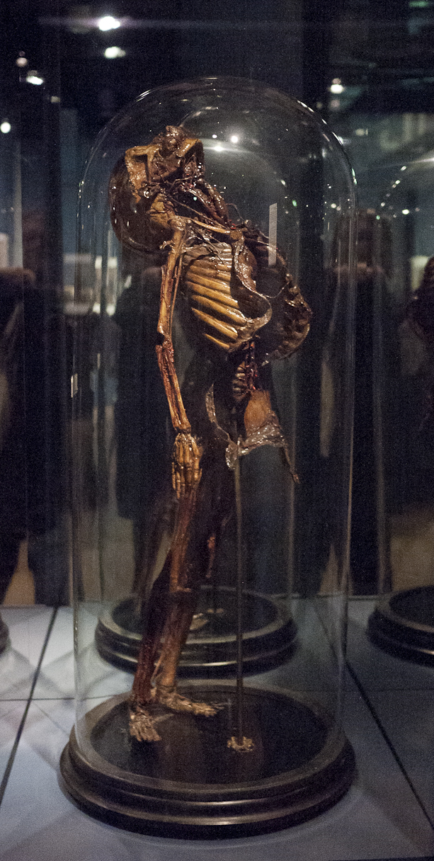
There were several "petrified" preparations in the collection (petrification being an Italian specialty), a fine osteological display, and a nice array of wet preparations. Of particular interest were the full-size flayed human specimens, whose vessels were injected with chalk (an odd method, but confirmed by several sources).

The mummy of Gaetano Arrighi, a convict who died in the early 19th century, seemed to have particular pride of place. His body went unclaimed and he was prepared according to a 19th century Italian recipe, but the results appear quite ancient. I fell in love with a particularly vivid, gesticulating infant in a nearby case, but the mummy certainly did have his charms.
You can find out more about the Museo di Anatomia Umana, Pisa (Museum of Human Anatomy at Pisa) by clicking here. You can read future posts by Evan both on this blog and on her Facebook page, which you will find by clicking here. All images are mine, from the museum.

Tuesday, November 27, 2012

The Vrolik Museum, Amsterdam, Re-designed and Re-opened!

I first visited the wonderful Amsterdam-based Vrolik Museum in 2007, when I was gathering material for the Anatomical Theatre, an exhibition exploring depictions of the body, disease, and death in medical museums of the Western world. The museum has has just reopened after a year-long closure for renovation and refurbishment; I asked friend and curator Dr. Laurens de Rooy to send in some photos and write a brief report about the museum and its redesign especially for the readers of this blog:
The Vrolik Museum is named after 19th Dutch anatomists Gerard (1775–1859) and Willem Vrolik (1801–1863). Their collection includes many zoological and comparative anatomical specimens, as well as many pathological specimens such as skeletons with rickets and other diseases of the bone. Since the redesign of the museum, these collections are now on display together for the first time since Willem Vrolik's death.
In the modern showcases you will find an overview of normal and abnormal anatomy-- e.g; a large series of the development of the brain--and of embryological development, siamese twins, cyclopes, development of the heart, dwarfism, anatomy of the limbs, corset livers etc etc. Along the walls is an overview of the long history of the collection, many of which displayed in original showcases like the 18th century Hovius cabinet (top image) and the 19th century Vroliks cabinet (images 2 and 3).
The Museum Vrolik is open on weekdays from 10 am to 5 pm. Entrance is free but we appreciate a voluntary admission of about 5 euros pp. All texts are both in English and Dutch. We provide a so called 'top-exhibits-tour' a little book with 30 highlights of the museum, including some congenital malformations, bezoar stones, bladder stones, corset livers, the lion that belonged to King Louis Napoleon, the skull of a man that got hit by a horse etc etc.
For more information about the museum, go to www.museumvrolik.nl. Via this site you may also book a guided tour.

Photos top to bottom:
  1. Hovius cabinet. cabinet from 1773 with pathologies of the bone, collected by physician Jacob Hovius (potrait on top of the cabinet)
  2. Vrolik's cabinet. Early 19th century cabinet of the old Museum Vrolikianum, showing the collection of comparative anatomy of father and son Vrolik (left portrait of Willem Vrolik and part of the collection of congenital malformations in animals; right: portrait of Gerard Vrolik and collection of human pathology and botany)
  3. Overview of the museum. Left and right: showcases with anatomy of head and neck and of skull, spine and thorax, in the center: a so called "blown skull" or "beauchene skull", all cranial bones have been taken apart and mounted separately; to the back; Vroliks cabinet
  4. The collection of animal skeletons and skulls of the original Vrolik collection, presented as a long chain or stairway from least perfect to most perfect. Father and son Vrolik did not believe in the possibility of evolution. All animals were created in their view according to a greater plan. The big animal skeleton top-left: the lion of king Louis Napoleon Bonaparte, king of Holland between 1806 and 1810.
  5. Human trunk with the heart and great vessels, modeled in red and blue wax.

Friday, October 5, 2012

"Ecstatic Raptures and Immaculate Corpses: Visions of Death Made Beautiful in Italy" Exhibition, Final Open Hours TOMORROW, Saturday October 6, Noon-7 PM


Tomorrow--Saturday, October 6--is your last chance to check out "Ecstatic Raptures and Immaculate Corpses: Visions of Death Made Beautiful in Italy," an exhibition featuring photographs by myself (some of which can be seen above) and waxworks by artists Eleanor Crook and Sigrid Sarda, on view at The Last Tuesday Society, 11 Mare Street, London All photographs and waxworks are for sale, and quite affordable, if I do say!

The exhibition will be view from Noon until 7:00 PM. Also on view will be the wonderful collection of taxidermy, naturalia, erotica, books and curiosities which comprises the spectacular Last Tuesday Society Giftshop.

Well worth a trip, I promise! Full details follow.
Ecstatic Raptures and Immaculate Corpses: Visions of Death Made Beautiful in Italy
An exhibition of photographs by Joanna Ebenstein of the Morbid Anatomy Blog, The Morbid Anatomy Library and Observatory with waxworks by Eleanor Crook and Sigrid Sarda.
Date: TOMORROW: Saturday, October 6
Time: Noon-7:00 PM
Location: The Last Tuesday Society, 11 Mare Street, London, E8 4RP
In her many projects, ranging from photography to curation to writing, New York based Joanna Ebenstein utilizes a combination of art and scholarship to tease out the ways in which the pre-rational roots of modernity are sublimated into ostensibly "purely rational" cultural activities such as science and medicine.Much of her work uses this approach to investigate historical moments or artifacts where art and science, death and beauty, spectacle and edification, faith and empiricism meet in ways that trouble contemporary categorical expectations.In the exhibition "Ecstatic Raptures and Immaculate Corpses" Ebenstein turns this approach to an examination of the uncanny and powerfully resonant representations of the dead, martyred, and anatomized body in Italy, monuments to humankind's quest to eternally preserve the corporeal body and defeat death in arenas sacred and profane.The artifacts she finds in both the churches, charnel houeses and anatomical museums of Italy complicate our ideas of the proper roles of--and divisions between--science and religion, death and beauty; art and science; eros and thanatos; sacred and profane; body and soul.
In this exhibition, you will be introduced to tantalizing visions of death made beautiful, uncanny monuments to the human dream of life eternal. You will meet "Blessed Ismelda Lambertini," an adolescent who fell into a fatal swoon of overwhelming joy at the moment of her first communion with Jesus Christ, now commemorated in a chillingly beautiful wax effigy in a Bolognese church; The Slashed Beauty, swooning with a grace at once spiritual and worldly as she makes a solemn offering of her immaculate viscera; Saint Vittoria, with slashed neck and golden ringlets, her waxen form reliquary to her own powerful bones; and the magnificent and troubling Anatomical Venuses, rapturously ecstatic life-sized wax women reclining voluptuously on silk and velvet cushions, asleep in their crystal coffins, awaiting animation by inquisitive hands eager to dissect them into their dozens of demountable, exactingly anatomically correct, wax parts.

Joanna Ebenstein
: New York based visual artist and independent scholar Joanna Ebenstein runs the popular Morbid Anatomy Blog and the related Morbid Anatomy Library, where her privately held collection of books, art, artifacts, and curiosities are made available by appointment.

For the past 5 years, she has traveled the world, seeking out the most curious, obscure and macabre collections, public and private, front stage and back, and sharing her findings via her the Morbid Anatomy Blog as well as a variety of exhibitions including  Anatomical Theatre, a photographic survey of artifacts of great medical museums of the Western World; The Secret Museum, a photographic exhibition exploring the poetics of collections private and public, front stage and back.

Other exhibitions using history as their muse include Savior of Mothers: The Forgotten Ballet of Dr. Ignaz Semmelweis  at the Center for Disease Control Museum and The Great Coney Island Spectacularium, an immersive investigation into the often bizarre spectacles of turn of the 20th century Coney Island at The Coney Island Museum.

She is the founding member of Observatory--a gallery and lecture space in Brooklyn, New York--and annual co-curator of The Congress for Curious Peoples, a 10-day series of lectures and performances investigating curiosity and curiosities, broadly considered and taking place at the Coney Island Museum.

Her work has been shown and published internationally, and she has lectured at museums and conferences around the world.
You can find out more about the show here, and view more images by clicking here.